***** '''Although magnesium ammonium phosphate and cystine stones are often radioopaque, they are not as dense as calcium oxalate or calcium phosphate stones'''
***** '''Although magnesium ammonium phosphate and cystine stones are often radioopaque, they are not as dense as calcium oxalate or calcium phosphate stones'''
**'''Nephrocalcinosis'''
***'''Formation of diffuse deposits of calcium throughout the kidneys'''
****'''Usually occurs within the renal medulla''' but occasionally it has been found in the cortex or within both the medulla and the cortex
****Minute calcifications seen in early stages may not be visible
***'''Can give rise to renal colic and hydronephrosis from dislodged calcific foci'''
***[[File:Nephrocalcinosis.jpg|alt=Nephrocalcinosis. Source: Wikipedia|thumb|Plain film x-ray demonstrating bilateral diffuse calcium deposits in the kidneys. Source: [[commons:File:Nephrocalcinosis.jpg|Wikipedia]]|400x400px]]'''Causes[https://radiopaedia.org/articles/medullary-nephrocalcinosis §]'''
****'''Medulla'''
*****'''Type 1 (distal) RTA'''
*****'''Hyperparathyroidism'''
*****'''Medullary sponge kidney'''
*****'''Hypervitaminosis D'''
*****'''Milk-alkali syndrome'''
*****'''Sarcoidosis'''
*****'''Hyper/hypothyroidism'''
*****'''Other pathological hypercalcemic or hypercalciuric states'''
***0.7 mSv with KUB[https://www.ncbi.nlm.nih.gov/pmc/articles/PMC5443345/ §]
**Cost (least expensive)
*'''Disadvantages'''
*'''Disadvantages'''
**'''Inability to visualize small stones'''
**'''Inability to visualize small stones'''
Line 31:
Line 65:
=== Ultrasound ===
=== Ultrasound ===
* '''Advantages'''
* Test characteristics[https://www.ncbi.nlm.nih.gov/pmc/articles/PMC5443345/ §]
**Sensitivity: 84%
**Specificity: 53%
*'''Advantages'''
**Availability
**Availability
**No radiation exposure
**No radiation exposure
**Cost
**Cost (5x cost of KUB)[https://www.ncbi.nlm.nih.gov/pmc/articles/PMC5443345/ §]
*'''Disadvantages (2):'''
*'''Disadvantages (2):'''
** '''Inability to visualize most ureteral stones'''
** '''Inability to visualize most ureteral stones'''
Line 43:
Line 80:
***US and CT measurements correlate 2/3 of the time
***US and CT measurements correlate 2/3 of the time
=== '''CT''' ===
=== CT scan (without contrast) ===
* '''Findings'''
* '''Findings'''
**'''Pure uric acid stones have much lower Hounsfield units than calcium types'''
**'''Pure uric acid stones have much lower Hounsfield units than calcium types'''
Line 49:
Line 86:
*** Usually associated with a small distal ureteral calculus.
*** Usually associated with a small distal ureteral calculus.
*** '''Should be similarly to other ureteral stones:''' intervention should be undertaken when there is an associated fever, nausea/vomiting, or unrelenting pain. Otherwise, conservative observation is appropriate.
*** '''Should be similarly to other ureteral stones:''' intervention should be undertaken when there is an associated fever, nausea/vomiting, or unrelenting pain. Otherwise, conservative observation is appropriate.
**Cardiac Lidocaine 1.5 mg/kg IV in 100 mL NS over 10 minutes (MAX 200 mg)
*'''<span style="color:#ff0000">Urologic Emergency: If obstructing stones with suspected infection, must urgently drain the collecting system with a stent or nephrostomy tube and delay stone treatment</span>[https://pubmed.ncbi.nlm.nih.gov/27238616/ ★]'''
**Definitive management of the stone should not be undertaken until sepsis has resolved and the infection has been treated with an appropriate course of antibiotic therapy.
== Diagnosis and Evaluation of Metabolic Stone Disease ==
== Diagnosis and Evaluation of Metabolic Stone Disease ==
* '''Goals of evaluation'''
=== UrologySchool.com Summary ===
**'''Identify potential associated metabolic disorders such as (5)'''
**** Surgical history should be obtained focusing particularly on bariatric surgery and surgeries of the intestinal tract. '''In contrast to gastric bypass surgery, restrictive bariatric surgeries such as gastric sleeve or gastric band do not seem to increase the risk for kidney stones'''
***'''<span style="color:#ff0000">Obtain or review available imaging studies to quantify stone burden.</span>'''
** '''<span style="color:#ff0000">One or two 24-hour urine collections</span>'''
**** Preoperative serum chemistries are important because they may provide clues to underlying serious diseases such as renal tubular acidosis or hypoparathyroidism or other metabolic derangements
***** Many infected calculi will harbour bacteria even after treatment with broad-spectrum antibiotics
***#'''<span style="color:#ff0000">Medical conditions predisposing to stones (e.g., RTA Type 1, primary hyperparathyroidism, gout, diabetes mellitus type)</span>'''
***** Half of infected calculi grow bacterial cultures that are different from the preoperative urine specimen
*** Urine microscopy for crystals may provide clues to diagnosis
****Insert urine microscopy table
***Stone composition, if available
****Can direct metabolic investigation or potentially obviate the need for a complete metabolic evaluation
* '''Extensive diagnostic evaluation'''
=== Goals of Evaluation ===
** Includes one or two 24-hour urine collections
*'''Identify potential associated metabolic disorders such as (5)'''
**Indications for a metabolic stone evaluation
*#'''Distal renal tubular acidosis (RTA)'''
**# Recurrent stone formers
*#'''Primary hyperparathyroidism'''
**# Strong family history of stones
*#'''Enteric hyperoxaluria'''
**# Intestinal disease
*#'''Cystinuria'''
**# Pathological skeletal fractures
*#'''Gouty diathesis'''
*'''Reduce risk of stone recurrence'''
**First-time stone formers have been estimated to have a 50% risk for recurrence within the subsequent 10 years
** Patients at higher risk for repeat episodes (6):
**# Family history of stones
**# Intestinal disease (particularly when causing chronic diarrheal states)
**# Pathologic skeletal fractures
**# Osteoporosis
**# Osteoporosis
**# History of UTI with calculi
**# UTI
**# Personal history of gout
**# Gout
**# Infirm health (unable to tolerate repeat stone episodes)
=== History and Physical Exam ===
**# Solitary kidney
**# Anatomic abnormalities
**# Stones composed of cystine, uric acid, and struvite
**# Children should generally be evaluated because of concerns about renal damage and long-term sequelae of stone recurrence
** '''Significant aberrations in total creatinine excretion from estimated volumes (males 20-25mg/kg and females 15-20mg/kg in 24 hours) imply incomplete collection, overcollection, greater than expected muscle mass, or less than expected muscle mass'''
*** For abnormally collected 24 hour urine collections, can divide metabolite excretion by creatinine excretion to compare collections
** The urinary constituents most commonly assayed in a 24 hours urine collection include calcium, oxalate, citrate, total volume, sodium, magnesium, potassium, pH, uric acid, and sulfate.
*** Sulfate is added to assess the volume of protein loading from animal meat.
== Acute management ==
==== History ====
* '''Screen for factors that predispose to calculi'''
** '''<span style="color:#ff0000">Conditions associated with stone disease (8):</span>'''
**#'''<span style="color:#ff0000">Malabsorptive gastrointestinal states</span>''' due to bowel resection, bariatric surgery or bowel or pancreatic disease
**##'''Chronic diarrhea that could be caused by inflammatory bowel disease (Crohn disease, ulcerative colitis) or irritable bowel syndrome'''
**## Gout may predispose the patient to hyperuricosuria or gouty diathesis with either uric acid calculi or calcium oxalate stone formers
**## '''Surgical history should be obtained focusing particularly on bariatric surgery and surgeries of the intestinal tract.'''
**###'''Roux-en-Y-gastric bypass surgery may significantly increase the overall risk for stone formation'''
**###'''In contrast to gastric bypass surgery, restrictive bariatric surgeries such as gastric sleeve or gastric band do not seem to increase the risk for kidney stones'''
***'''Should include average daily intake of fluids (amount and specific beverages), protein (types and amounts), calcium, sodium, high oxalate-containing foods, fruits and vegetables and over-the-counter supplements.'''
**** '''Nutritional factors associated with stone disease, depending on stone type and risk factors, include'''
*****Calcium intake below or significantly above the recommended dietary allowance (RDA)
** Cardiac Lidocaine 1.5 mg/kg IV in 100 mL NS over 10 minutes (MAX 200 mg)
** Acetaminophen 1000 mg PO
** 1 L 0.9% NS bolus
== Conservative management ==
* '''Body mass index'''
* '''Fluid recommendations'''
** '''Increase fluid intake to achieve a daily urine output of ≥2 liters'''
*** '''Overall, most evidence suggests that it is not the type of fluid ingested that is important for stone prevention but rather the absolute amount of fluid volume taken in per day'''
**** Water hardness should be a minor concern with respect to stone formation
**** Carbonated water may provide some protective benefit
**** Citrus juices (particularly lemon and orange juices) may be a useful adjunct to stone prevention
**** '''Soda flavored with phosphoric acid may increase stone risk, whereas those with citric acid may decrease risk'''
***** Several sodas are acidified by citric acid and contain an amount of citrate equal to or greater than that of lemonade, including Diet Sunkist Orange, Diet 7Up, Sprite Zero, Diet Canada Dry Ginger Ale, Sierra Mist Free, Diet Orange Crush, Fresca, and Diet Mountain Dew. All of the aforementioned sodas have the potential to decrease the risk of kidney stones similar to or greater than lemonade.
***** In contrast, colas, including Caffeine Free Diet Coke, Diet Coke, Diet Coke with Lime, Coke Zero, Caffeine Free Diet Pepsi and Pepsi, are acidified by phosphoric acid, not by citric acid and contain low citrate levels.
****** One randomized study of recurrent stone formers with baseline soda consumption > 160 ml per day, found that over a 3-year period those who abstained from any soft drink consumption had a lower risk of symptomatic stone events (34%) compared to those who continued to drink sodas acidified by phosphoric acid (41%; RR, 0.83).§
**** '''Performance sports drinks'''
***** '''May increase urinary citrate and pH''' thereby reducing risk of stones.
****** However, these drinks have a high fructose and total carbohydrate content so they should not be recommended as the primary means of hydration for stone formers.
***** Do not lead to hypernatriuria, even though sodium content may be high
***** No effect on urinary calcium, oxalate, and uric acid.
*** RCTs have demonstrated a benefit of a diet with reduced animal protein (meat) intake
** '''Sodium restriction'''
*** '''High sodium intake is associated with:'''
***# '''Increases calcium excretion'''
***# '''Increases urinary pH'''
***# '''Decreases citrate excretion'''
*** RCTs have demonstrated a benefit of dietary sodium restriction
** Weight-loss diets
*** The consumption of a low-carbohydrate, high-protein diet delivers a marked acid load to the kidney, increases the risk for stone formation, and may increase the risk of bone loss
* '''Obesity'''
** '''Increased BMI, larger waist size, and weight gain are correlated with an increased risk for stone episodes'''
** '''Increased BMI, larger waist size, and weight gain are correlated with an increased risk for stone episodes'''
*** '''The association of obesity and uric acid stone formation is primarily due to change in urinary pH'''
*** '''The association of obesity and uric acid stone formation is primarily due to change in urinary pH'''
*** '''The association of obesity with calcium oxalate stone formation is primarily due to increased excretion of promoters of stone formation''' (oxalate, uric acid, sodium, and phosphorus)
*** '''The association of obesity with calcium oxalate stone formation is primarily due to increased excretion of promoters of stone formation''' (oxalate, uric acid, sodium, and phosphorus)
** '''Metabolic syndrome is associated with lower urinary pH'''
=== Laboratory ===
** Roux-en-Y-gastric bypass surgery may significantly increase the overall risk for stone formation
* '''<span style="color:#ff0000">Urinalysis should include pH</span>'''
*** '''Restricted calcium intake leads to an increase in available intestinal oxalate, which subsequently increases oxalate absorption,''' thereby raising the supersaturation of calcium oxalate;
** '''<span style="color:#ff0000">pH > 7.0 is suggestive of infection lithiasis or RTA</span>'''
** '''Calcium supplements are safe if attention is paid to preparation (calcium citrate appears to be a more stone-friendly calcium supplement because of the additional inhibitory action of citrate) and especially timing (should be taken with meals)'''
* May suggest underlying medical conditions associated with stone disease (e.g., primary hyperparathyroidism, gout, RTA type 1 or other metabolic derangements)
* '''Assessment of underlying renal function is necessary'''
**#'''<span style="color:#ff0000">Predominantly calcium phosphate stone composition</span>'''
*'''Measurement of vitamin D levels may be helpful as''' '''low vitamin D levels may mask primary hyperparathyroidism, or contribute to secondary hyperparathyroidism.'''
*A high or high normal intact PTH in these settings should prompt further endocrine evaluation, imaging or referral for consideration of parathyroidectomy.
==== Stone composition, if available ====
*'''When a stone is available, a stone analysis should be obtained at least once.'''
*Can direct metabolic investigation or potentially obviate the need for a complete metabolic evaluation
*'''<span style="color:#ff0000">Calcium phosphate stone composition associated with:</span>'''
*#'''<span style="color:#ff0000">RTA Type 1</span>'''
*#'''<span style="color:#ff0000">Use of carbonic anhydrase inhibitors</span>'''
=== Imaging ===
* '''Obtain or review available imaging studies to quantify stone burden.''' '''<span style="color:#ff0000">[https://pubmed.ncbi.nlm.nih.gov/24857648/ ★]</span>'''
=== Metabolic/Extended Diagnostic Evaluation ===
* '''<span style="color:#ff0000">Consists of one or two 24-hour urine collections obtained on a random diet[https://pubmed.ncbi.nlm.nih.gov/24857648/ ★]</span>'''
*#'''In stone formers with known cystine stones or a family history of cystinuria or for those in whom cystinuria is suspected, urinary cystine should additionally be measured.'''
*#Sulfate can be added to assess the volume of protein loading from animal meat
* '''<span style="color:#ff0000">Assess adequacy of 24-hour urine collection, prior to interpretation of results'''
**'''<span style="color:#ff0000">To assess the adequacy of collection, 24-hour urinary creatinine excretion should be considered</span>, taking into account patient gender and body weight (males 20-25mg/kg and females 15-20mg/kg in 24 hours),''' as well as patient recall of the start and end times of his or her collection
***'''Significant aberrations in total creatinine excretion from estimated volumes imply incomplete collection, overcollection, greater than expected muscle mass, or less than expected muscle mass'''
**** For abnormally collected 24 hour urine collections, can divide metabolite excretion by creatinine excretion to compare collections
*'''Markers of protein intake, such as urine urea nitrogen or urinary sulfate, are reflective of animal protein intake and can be used to assess dietary adherence'''.
*Urinary potassium measured at baseline can be compared to urinary potassium obtained during follow-up to gauge compliance with medication regimens.
*'''Primary hyperoxaluria should be suspected when urinary oxalate excretion > 75 mg/day in adults without bowel dysfunction. These patients should be considered for referral for genetic testing and/or specialized urine testing'''
*Fast and calcium load testing should not be performed routinely to distinguish among types of hypercalciuria
*If a patient with calcium urolithiasis uses calcium supplements, 24-hour urine samples should be collected on and off the supplement.
**If urinary supersaturation of the calcium salt in question increases during the period of supplement use, the supplement should be discontinued.
== Diet Therapies ==
* '''<span style="color:#ff0000">General diet therapies to reduce risk of stone recurrence (6)'''
*# '''<span style="color:#ff0000">Moderate calcium intake (1,000-1,200 mg per day)'''
*# '''<span style="color:#ff0000">Limit intake of oxalate-rich foods'''
*# '''<span style="color:#ff0000">Increase intake of fruits and vegetables'''
*# '''<span style="color:#ff0000">Limit intake of non-dairy animal protein'''
=== Increase fluid intake ===
#'''<span style="color:#ff0000">Fluid intake that will achieve a urine volume of > 2.5 liters daily is recommended in all stone formers</span>'''
#*An RCT of recurrent calcium oxalate stone formers randomized to a high fluid intake vs. no specific recommendations found significantly reduced stone recurrence rates in the high fluid intake group (12% vs. 27%, respectively, at 5 years)
#*Although there is no definitive threshold for urine volume and increased risk, an accepted goal is ≥2.5 liters of urine daily.#**Because of insensible losses and varying intake of fluid contained in food, a universal recommendation for total fluid intake is not appropriate
#*'''Overall, most evidence suggests that it is not the type of fluid ingested that is important for stone prevention but rather the absolute amount of fluid volume taken in per day'''
#** Water hardness should be a minor concern with respect to stone formation
#** Carbonated water may provide some protective benefit
#** Citrus juices (particularly lemon and orange juices) may be a useful adjunct to stone prevention
#**'''Alcoholic beverages, coffee, decaffeinated coffee, tea and wine have been shown to be associated with a lower risk of stone formation'''
#**'''Sugar-sweetened beverages demonstrated an increased risk.'''
#***'''The only specific beverage that has been evaluated for an effect on stone recurrence in an RCT is soft drinks; the group avoiding soft drinks demonstrated a marginally lower rate of stone recurrence at the end of the 3-year trial but the effect appeared to be limited to those consuming primarily phosphoric acid-based (e.g. colas) rather than citric acid-based soft drinks'''
#***'''Soda flavored with phosphoric acid may increase stone risk, whereas those with citric acid may decrease risk'''
#**** Several sodas are acidified by citric acid and contain an amount of citrate equal to or greater than that of lemonade, including Diet Sunkist Orange, Diet 7Up, Sprite Zero, Diet Canada Dry Ginger Ale, Sierra Mist Free, Diet Orange Crush, Fresca, and Diet Mountain Dew. All of the aforementioned sodas have the potential to decrease the risk of kidney stones similar to or greater than lemonade.
#**** In contrast, colas, including Caffeine Free Diet Coke, Diet Coke, Diet Coke with Lime, Coke Zero, Caffeine Free Diet Pepsi and Pepsi, are acidified by phosphoric acid, not by citric acid and contain low citrate levels.
#***** One randomized study of recurrent stone formers with baseline soda consumption > 160 ml per day, found that over a 3-year period those who abstained from any soft drink consumption had a lower risk of symptomatic stone events (34%) compared to those who continued to drink sodas acidified by phosphoric acid (41%; RR, 0.83).§
#***'''Performance sports drinks'''
#**** '''May increase urinary citrate and pH''' thereby reducing risk of stones.
#***** However, these drinks have a high fructose and total carbohydrate content so they should not be recommended as the primary means of hydration for stone formers.
#**** Do not lead to hypernatriuria, even though sodium content may be high
#**** No effect on urinary calcium, oxalate, and uric acid.
=== Limiting sodium intake ===
* '''<span style="color:#ff0000">Limiting sodium intake (target of ≤100 mEq (2,300 mg)) is recommended in patients with calcium stones and relatively high urinary calcium</span>'''
* '''High sodium intake is associated with:'''
*# '''Increased calcium excretion'''
*# '''Increased urinary pH'''
*# '''Decreased citrate excretion'''
* RCTs have demonstrated a benefit of dietary sodium restriction
=== Moderate calcium intake ===
* '''<span style="color:#ff0000">Consuming 1,000-1,200 mg per day of dietary calcium is recommended in patients with calcium stones and relatively high urinary calcium</span>'''
**'''A lower calcium diet in the absence of other specific dietary measures is associated with an increased risk of stone formation'''
***'''Lower calcium intake results in insufficient calcium to bind dietary oxalate in the gut, thereby increasing oxalate absorption and urinary oxalate excretion.'''
**'''In contrast, the RDA of calcium, defined as 1,000-1,200 mg/day for most individuals, was shown to be associated with reduced risk'''
* '''Calcium supplements are safe if attention is paid to preparation (calcium citrate appears to be a more stone-friendly calcium supplement because of the additional inhibitory action of citrate) and especially timing (should be taken with meals)'''
=== Limit intake of oxalate-rich foods ===
* '''<span style="color:#ff0000">Limiting intake of oxalate-rich foods and maintaining normal calcium consumption is recommended in patients with calcium oxalate stones and relatively high urinary oxalate</span>'''
*'''Urinary oxalate is also modulated by calcium intake, which influences intestinal oxalate absorption'''
*'''Other factors that may contribute to higher urinary oxalate include vitamin C''' (ascorbic acid is metabolized to oxalate) '''and other over-the-counter nutrition supplements.'''
*Although dietary oxalate clearly plays a role in increased urinary oxalate, it is difficult to restrict its intake because oxalate is ubiquitous and found in most vegetable matter. However, it is important to avoid large portions of '''foodstuffs that are rich in oxalate, such as spinach, beets, chocolate, nuts, and tea.'''
* Whereas general advice on a restricted-oxalate intake might be given to patients with recurrent nephrolithiasis, '''a low-oxalate diet would be most useful in patients with enteric hyperoxaluria, such as those with underlying bowel abnormalities or previous gastric bypass surgery'''
=== Increase intake of fruits and vegetables ===
* '''<span style="color:#ff0000">Recommended in patients with calcium stones and relatively low urinary citrate</span>'''
**Although a number of fruits and juices have been evaluated for their effect on urinary stone risk factors, none have been prospectively evaluated in an RCT assessing actual stone formation.
*'''Urinary citrate excretion is determined by acid-base status; conditions such as metabolic acidosis, renal tubular acidosis and chronic diarrhea, and some medications, such as carbonic anhydrase inhibitors, may promote hypocitraturia'''
**Acidosis can arise from a diet that is inordinately rich in foods with a high potential renal acid load such as meats, fish, poultry, cheese, eggs, and to a lesser extent, grains.
=== Limit intake of non-dairy animal protein ===
* '''<span style="color:#ff0000">Recommended in patients with calcium stones and relatively low urinary citrate</span>'''
* '''<span style="color:#ff0000">May help reduce stone recurrence in patients with uric acid stones or calcium stones and relatively high urinary uric acid</span>'''
*'''Protein intake increases urinary calcium, oxalate, and uric acid excretion'''
**'''Urinary uric acid is derived from both endogenous and exogenous sources'''
***Diet-derived purines account for an ≈30% of urinary uric acid
* '''If diet assessment suggests that purine intake is contributory to high urinary uric acid, patients may benefit from limiting high- and moderately high purine containing foods.'''
**'''"High purine" foods are generally considered specific fish and seafood (anchovies, sardines,''' herring, mackerel, '''scallops and mussels)''', water fowl, '''organ meats,''' glandular tissue, gravies and meat extracts.
**'''"Moderately-high" sources of purines include other shellfish and fish,''' game meats, mutton, '''beef, pork, poultry''' and meat-based soups and broths
*RCTs have demonstrated a benefit of a diet with reduced animal protein (meat) intake
=== Cystine Stones ===
*'''Patients with cystine stones should be counselled to increase fluid intake and limit sodium and protein intake'''
**'''High fluid intake is particularly important in cystine stone formers'''; the target for urine volume is typically higher than that recommended to other stone formers; '''oral intake of ≥4 L/day is often required'''
**Lower sodium intake has been shown to reduce cystine excretion
**Limiting animal protein intake is of benefit in patients with cystine stones
=== Other ===
*Weight-loss diets
** The consumption of a low-carbohydrate, high-protein diet delivers a marked acid load to the kidney, increases the risk for stone formation, and may increase the risk of bone loss
* '''Vitamin D'''
* '''Vitamin D'''
** '''Controversy exists over the role of vitamin D supplementation and kidney stone formation'''
** '''Controversy exists over the role of vitamin D supplementation and kidney stone formation'''
** '''Patients who undergo vitamin D repletion should have their 24-hour urine calcium monitored'''
** '''Patients who undergo vitamin D repletion should have their 24-hour urine calcium monitored'''
* '''Dietary oxalate'''
** Although dietary oxalate clearly plays a role in increased urinary oxalate, it is difficult to restrict its intake because oxalate is ubiquitous and found in most vegetable matter. However, it is important to avoid large portions of foodstuffs that are rich in oxalate, such as spinach, beets, chocolate, nuts, and tea.
** Whereas general advice on a restricted-oxalate intake might be given to patients with recurrent nephrolithiasis, '''a low-oxalate diet would be most useful in patients with enteric hyperoxaluria, such as those with underlying bowel abnormalities or previous gastric bypass surgery'''
* Follow-up
** If the patient’s metabolic or environmental abnormalities have been corrected, the conservative therapy can be continued and the patient followed every 6-12 months with repeat 24-hour urine testing as indicated.
*** Follow-up is essential not only to monitor the efficiency of treatment but also to encourage patient compliance. If, however, a metabolic defect persists, a more selective medical therapy may be instituted.
== Selective medical therapy for nephrolithiasis ==
*** Recall, in absorptive hypercalciuria I, increased absorption will occur regardless of the amount of calcium in the patient’s diet (these patients will demonstrate an hypercalcuria on both the fasting and the loading specimens). In contrast, patients with absorptive hypercalciuria II will have a normal amount of urinary calcium excretion during calcium restriction, but will show elevations during their regular diet
***'''<span style="color:#ff0000">Recurrent calcium or calcium phosphate stones</span>'''
*** Currently, no treatment is capable of correcting the basic abnormality of type I absorptive hypercalcuria
* '''Mechanism of Action'''
*** Thiazides may have a limited long-term effectiveness in type I absorptive hypercalcuria
*# '''Directly stimulate calcium reabsorption in the distal nephron''' (hypocalcuric effect)
*** In type II absorptive hypercalciuria, no specific drug treatment may be necessary because the physiologic defect is not as severe as in type I.
*# '''Promotes excretion of sodium causing extracellular volume depletion'''
** '''Thiazides'''
*#* '''Long-term thiazide therapy results in volume depletion, extracellular volume contraction, and proximal tubular resorption of sodium and calcium.'''
*** '''MOA'''
* '''<span style="color:#ff0000">Drugs and dosages'''
***# '''Directly stimulate calcium reabsorption in the distal nephron'''
*#'''<span style="color:#ff0000">Hydrochlorothiazide</span> (50mg orally, once daily;''' 25mg orally, twice daily''')'''
***# '''Promotes excretion of sodium causing extracellular volume depletion'''
*#'''<span style="color:#ff0000">Chlorthalidone</span>''' (25mg orally, once daily)
***** '''Long-term thiazide therapy results in volume depletion, extracellular volume contraction, and proximal tubular resorption of sodium and calcium.'''
*#'''<span style="color:#ff0000">Indapamide</span> (2.5mg orally, once daily)'''
*** Chlorthalidone (25-50 mg/day) or indapamide (2.5 mg/day) are preferred to hydrochlorothiazide since they are long-acting and are once a day dosing.
*#*Chlorthalidone or indapamide are preferred to hydrochlorothiazide since they are long-acting and are once a day dosing.
**** Indapamide is technically not a thiazide but does share a successful hypocalciuric effect with the other agents.
*#* Indapamide is technically not a thiazide but does share a successful hypocalciuric effect with the other agents.
*** '''Patients placed on thiazide diuretics for management of hypercalciuria should also be placed on dietary sodium restriction'''
* '''<span style="color:#ff0000">Patients placed on thiazide diuretics for management of hypercalciuria should also be placed on dietary sodium restriction'''
**** An excess sodium load will inhibit reabsorption of calcium in the proximal tubule, thereby causing hypercalciuria.
** '''An excess sodium load will inhibit reabsorption of calcium in the proximal tubule, thereby causing hypercalciuria.'''
***'''<span style="color:#ff0000">Potassium supplementation (either potassium citrate or potassium chloride)</span>'''
****'''<span style="color:#ff0000">May be needed when thiazide therapy is employed because of the hypokalemic effects of these medications</span>'''
****'''<span style="color:#ff0000">Should always be considered at either the onset of thiazide therapy or upon the discovery of glucose intolerance'''
***** '''Can prevent or significantly lessens the degree of hypokalemia or glucose intolerance'''
***** '''Can be administered as potassium citrate or with dietary supplements such as a banana per day'''
****** '''Citrates are generally well tolerated, with only a small risk for GI upset'''
******* A liquid preparation of potassium citrate, rather than the slow-release tablet preparation, is recommended in patients with rapid intestinal transit time (i.e. chronic diarrhea); the slow-release medication may be poorly absorbed
****** Conflicting evidence of an increased risk of calcium phosphate stone formation with the long-term use of potassium citrate therapy
***** Particularly important in patients with evident potassium deficiency, patients on digitalis therapy, and those individuals who develop hypocitraturia
****'''The addition of amiloride or spironolactone may avoid the need for potassium supplementation'''.
****Triamterene, although it is potassium-sparing, should be avoided as stones of this compound have been reported
**'''Hypocitraturia'''
***'''Result of hypokalemia with intracellular acidosis'''
**'''Patients with undiagnosed primary hyperparathyroidism may develop hypercalcemia after initiation of thiazide therapy'''
*** Although most patients with primary hyperparathyroidism demonstrate hypercalcemia and hypercalciuria, a normal serum calcium level in the presence of an inappropriately high serum PTH value may be seen in some cases, making the diagnosis more difficult. Administration of a thiazide diuretic will enhance renal calcium reabsorption and exacerbate the hypercalcemia, thereby facilitating the diagnosis (“thiazide challenge”)
**** In the thiazide challenge, cessation of thiazide should reduce PTH and serum calcium. However, in primary hyperparathyroidism, PTH and serum calcium remain persistently elevated with cessation of thiazide.
** '''Bisphosphonates combined with thiazide diuretics appear to reduce hypercalciuria while protecting the bone'''
* '''Thiazide diuretics lose their effectiveness in the treatment of hypercalciuria in up to 25% of patients on long-term management.'''
** '''Loss of effectiveness is due to increased serum calcium levels which stimulate the C cells in the thyroid to produce more calcitonin.''' Increased calcitonin leads to increased urinary calcium excretion.
*** '''Can prevent or significantly lessens the degree of hypokalemia or glucose intolerance'''
**#'''<span style="color:#ff0000">Recurrent calcium or calcium phosphate stones</span>'''
*** '''Can be administered as potassium citrate, potassium-sparing agents (triamterene), or with dietary supplements such as a banana per day'''
**#*'''Citrates are first-line therapy for the management of RTA, thiazide-induced hypocitraturia, and idiopathic hypocitraturia'''
**** '''Citrates are generally well tolerated, with only a small risk for GI upset'''
**#** Potassium citrate therapy is able to correct the metabolic acidosis and hypokalemia found in patients with distal RTA
***** A liquid preparation of potassium citrate, rather than the slow-release tablet preparation, is recommended in patients with rapid intestinal transit time (i.e. chronic diarrhea); the slow-release medication may be poorly absorbed
**#*'''Calcium stone-forming patients with normal citrate excretion but low urinary pH may also benefit from citrate therapy'''
**** Conflicting evidence of an increased risk of calcium phosphate stone formation with the long-term use of potassium citrate therapy
**#**There is also a risk that higher urine pH can promote calcium phosphate stone formation, or change calcium oxalate stone formers to calcium phosphate stone formers.
*** Particularly important in patients with evident potassium deficiency, patients on digitalis therapy, and those individuals who develop hypocitraturia
**#* '''Potassium citrate is preferred over sodium citrate'''
* '''Hypocitraturia'''
**#**'''Patient's treated with sodium alkali will occasionally begin forming calcium oxalate stones due to an excess sodium load that will inhibit reabsorption of calcium in the proximal tubule, thereby causing hypercalciuria'''
** '''Result of hypokalemia with intracellular acidosis'''
**#**'''If the patient is at risk for hyperkalemia, other agents such as sodium bicarbonate or sodium citrate should be considered.'''
**** '''Patients with undiagnosed primary hyperparathyroidism may develop hypercalcemia after initiation of thiazide therapy'''
***** Although most patients with primary hyperparathyroidism demonstrate hypercalcemia and hypercalciuria, a normal serum calcium level in the presence of an inappropriately high serum PTH value may be seen in some cases, making the diagnosis more difficult. Administration of a thiazide diuretic will enhance renal calcium reabsorption and exacerbate the hypercalcemia, thereby facilitating the diagnosis (“thiazide challenge”)
****** In the thiazide challenge, cessation of thiazide should reduce PTH and serum calcium. However, in primary hyperparathyroidism, PTH and serum calcium remain persistently elevated with cessation of thiazide.
**** '''Bisphosphonates combined with thiazide diuretics appear to reduce hypercalciuria while protecting the bone'''
*** '''Thiazide diuretics lose their effectiveness in the treatment of hypercalciuria in up to 25% of patients on long-term management.'''
**** '''Loss of effectiveness is due to increased serum calcium levels which stimulate the C cells in the thyroid to produce more calcitonin.''' Increased calcitonin leads to increased urinary calcium excretion.
** '''Fish oil'''
*** '''An effective, first-line therapy for mild-moderate hypercalciuria'''
**** Thought to have a protective role in preventing nephrolithiasis by decreasing urinary calcium and oxalate excretion through alteration of prostaglandin metabolism
** '''Hyperparathyroidism complicated by stone disease is best treated with surgical excision of the adenoma'''
** '''Patients with hyperuricosuria should be instructed to decrease dietary purine intake'''
*'''<span style="color:#ff0000">Allopurinol should be offered to patients with recurrent calcium oxalate stones who have hyperuricosuria and normal urinary calcium</span>'''
** '''Pharmacologic approaches to the management of hyperuricosuric calcium nephrolithiasis (2):'''
**'''Hyperuricemia is not a required criterion for allopurinol therapy'''
**# '''Alkalinizing the urine so that uric acid remains in a dissolved state'''
**'''In addition to medication, patients with hyperuricosuria should be instructed to limit non-dairy animal protein, which also may maximize the efficacy of allopurinol.'''
**#* '''Potassium citrate may be used'''
***Allopurinol’s use in hyperuricosuria associated with dietary purine overindulgence also may be reasonable if patients are unable or unwilling to comply with dietary purine restriction.
**#** CUA Guidelines only discuss allopurinol for hyperuricosuric calcium oxalate nephrolithiasis, not potassium citrate
*'''<span style="color:#ff0000">Thiazide diuretics and/or potassium citrate should be offered to patients with recurrent calcium stones in whom other metabolic abnormalities are absent or have been appropriately addressed and stone formation persists</span>'''
**# '''Decreasing the production of uric acid'''
**Both thiazides and potassium citrate therapy have been shown to prevent recurrent stones in patients with normal range urinary calcium and citrate, respectively
**** '''Allopurinol (300 mg/day) may be used'''
**'''For patients with no identified risk factors for nephrolithiasis, potassium citrate may be the preferred first-line therapy, given its relatively low side effect profile.'''
***** MOA: blocks the ability of xanthine oxidase to convert xanthine to uric acid.
===Uric acid stones===
****** The resultant decrease in serum uric acid will ultimately lead to a decrease in urinary uric acid as well.
*'''<span style="color:#ff0000">First-line therapy for patients with uric acid stones is alkalinization of the urine with potassium citrate to raise urinary pH to an optimal level so that uric acid remains in a dissolved state.</span>'''
***** Allopurinol’s use in hyperuricosuria associated with dietary purine overindulgence also may be reasonable if patients are unable or unwilling to comply with dietary purine restriction.
**'''<span style="color:#ff0000">Allopurinol should not be routinely offered as first-line therapy to patients with uric acid stones</span>'''
***'''Most patients with uric acid stones have low urinary pH rather than hyperuricosuria as the predominant risk factor'''
***'''<span style="color:#ff0000">Goal is to increase the urinary pH > 5.5 (AUA targets 6.0 and CUA targets 6.5), through an alkalinizing agent such as potassium citrate'''
**** With adequate alkali therapy, patient's can raise the urine pH to an optimal level so that uric acid remains in a dissolved state.
***** '''<span style="color:#ff0000">Attempts at alkalinizing the urine to a pH > 7.0 should be avoided. At a higher pH, there is a danger of increasing the risk for calcium phosphate stone formation.'''
***Patients may initially present with low/normal 24-hour urinary uric acid levels because the uric acid will precipitate out of solution in the acid urinary environment. Once the urine has been alkalized, all of the uric acid will come back into solution, causing a significant increase in the measured urinary uric acid.
*'''<span style="color:#ff0000">Allopurinol may be considered as an adjunct when alkalinization is not successful or for patients who continue to form uric acid stones despite adequate alkalinization of the urine.</span>'''
**'''Allopurinol (300 mg/day) may be used'''
*** MOA: blocks the ability of xanthine oxidase to convert xanthine to uric acid, resulting in decreased production of uric acid
**** The resultant decrease in serum uric acid will ultimately lead to a decrease in urinary uric acid as well.
* '''Acetazolamide is effective in increasing the urinary pH in patients with uric acid and cystine stone formation who are already taking potassium citrate.'''
** Acetazolamide, a carbonic anhydrase inhibitor, leads to an increase in urinary bicarbonate and increased H+ reabsorption.
** Up to 50% of patients may discontinue acetazolamide due to adverse effects.
===Uric acid and cystine stones===
* '''<span style="color:#ff0000">Potassium citrate should be offered to patients with uric acid and cystine stones to raise urinary pH to an optimal level</span>'''
**'''For uric acid stone formers, a urine pH of 6.0 (CUA targets 6.5)''' '''should be achieved'''
**'''For cystine stone formers, a urine pH of 7.0 (CUA targets >7.0) should be achieved'''
===Cystine stones===
*'''<span style="color:#ff0000">First-line therapy for patients with cystine stones:</span>'''
*#'''<span style="color:#ff0000">Restriction of sodium and protein intake</span>'''
*##Excess dietary sodium can lead to increases in cystine excretion
*'''<span style="color:#ff0000">Potassium citrate should be offered to patients with cystine stones to raise urinary pH to an optimal level</span>'''
**'''AUA: Urine pH of 7.0 (CUA targets >7.0) should be achieved'''
*'''<span style="color:#ff0000">Cystine-binding thiol drugs, such as alpha-mercaptopropionylglycine (tiopronin), should be offered to patients with cystine stones who are unresponsive to dietary modifications and urinary alkalinization, or have large recurrent stone burdens.</span>'''
** '''MOA: increase cystine solubility in urine by formation of a more soluble mixed-disulfide bond (i.e., cystine to drug, rather than cystine to cystine).'''
** '''Options include α-mercaptopropionylglycine (tiopronin [Thiola]),''' D-penicillamine (Cuprimine), and captopril
*** '''Tiopronin is possibly more effective and associated with fewer adverse events than d-penicillamine and should be considered first.'''
***'''Captopril, another thiol agent, has not been shown to be effective for the prevention of recurrent cystine stones'''
***d-Penicillamine and α-MPG are equally effective in their ability to decrease urinary cystine levels. However, α-mercaptopropionylglycine is significantly less toxic than d-penicillamine.
*** Side effects of '''D-penicillamine''' include gastrointestinal disturbances, fever and rash, arthralgia, leukopenia, thrombocytopenia, proteinuria with nephrotic syndrome, polymyositis, and '''pyridoxine (Vitamin B6) deficiency'''
**** '''Pyridoxine (vitamin B6) deficiency supplementation is recommended'''
**'''The medical management of infection calculi centers on the prevention of recurrence, rather than medical dissolution.'''
*'''<span style="color:#ff0000">Acetohydroxamic acid (AHA) may be offered to patients with residual or recurrent struvite stones only after surgical options have been exhausted.</span>'''
** '''Patients treated for struvite stones may still be at risk for recurrent UTIs after stone removal, and in some patients surgical stone removal is not feasible.'''
**'''The use of a urease inhibitor, AHA, may be beneficial in these patients, although the extensive side effect profile may limit its use. In particular, patients taking this medication should be closely monitored for phlebitis and hypercoagulable phenomena'''
***** '''Occurs in up to 15% of the patients; more prevalent in patients with renal insufficiency'''
****'''<span style="color:#ff0000">Phlebitis'''
*** '''Frequently reserved for patients deemed too ill for surgical management.'''
*Long-standing effective control of infection with urea-splitting organisms should be achieved if at all possible with improved bladder health, adequate urinary drainage, and suppressive antibiotics
*'''Phosphate therapy is contraindicated in cases of infection calculi because this medication may promote further stone formation.'''
=== Other ===
* '''Fish oil'''
** '''An effective, first-line therapy for mild-moderate hypercalciuria'''
*** Thought to have a protective role in preventing nephrolithiasis by decreasing urinary calcium and oxalate excretion through alteration of prostaglandin metabolism
* '''Hyperparathyroidism complicated by stone disease'''
**'''Best treated with surgical excision of the adenoma'''
* '''Enteric hyperoxaluria'''
* '''Enteric hyperoxaluria'''
Line 251:
Line 556:
** '''Dietary/supplemental calcium may help bind intestinal oxalate and decrease its absorption'''
** '''Dietary/supplemental calcium may help bind intestinal oxalate and decrease its absorption'''
** '''Treatment with potassium citrate (60 to 120 mEq/day) may correct the hypokalemia and metabolic acidosis in patients with enteric hyperoxaluria''' and, in some individuals, increase urinary citrate toward normal.
** '''Treatment with potassium citrate (60 to 120 mEq/day) may correct the hypokalemia and metabolic acidosis in patients with enteric hyperoxaluria''' and, in some individuals, increase urinary citrate toward normal.
** '''Citrates are first-line therapy for the management of RTA, thiazide-induced hypocitraturia, and idiopathic hypocitraturia'''
*** Potassium citrate therapy is able to correct the metabolic acidosis and hypokalemia found in patients with distal RTA
* '''Hypomagnesuric Calcium Nephrolithiasis'''
* '''Hypomagnesuric Calcium Nephrolithiasis'''
Line 263:
Line 562:
*** Potassium-magnesium may restore urinary magnesium and citrate levels with minimal GI side effects
*** Potassium-magnesium may restore urinary magnesium and citrate levels with minimal GI side effects
* '''Uric acid stones'''
=== Medical Management of Pediatric Calculi ===
** '''Goal is to increase the urinary pH > 5.5, preferably between 6.0-6.5, through an alkalinizing agent such as potassium citrate'''
*** With adequate alkali therapy, patient's can raise the urine pH above the dissociation constant of uric acid.
**** '''Attempts at alkalinizing the urine to a pH > 7.0 should be avoided. At a higher pH, there is a danger of increasing the risk for calcium phosphate stone formation.'''
*** Patients may initially present with low/normal 24-hour urinary uric acid levels because the uric acid will precipitate out of solution in the acid urinary environment. Once the urine has been alkalized, all of the uric acid will come back into solution, causing a significant increase in the measured urinary uric acid.
** '''Patient's treated with sodium alkali will occasionally begin forming calcium oxalate stones due to an excess sodium load that will inhibit reabsorption of calcium in the proximal tubule, thereby causing hypercalciuria'''
*** Potassium citrate is advantageous because it is not only a good alkalinizing agent, but it appears to be devoid of the complication of calcium stones.
** '''Acetazolamide is effective in increasing the urinary pH in patients with uric acid and cystine stone formation who are already taking potassium citrate.'''
*** Acetazolamide, a carbonic anhydrase inhibitor, leads to an increase in urinary bicarbonate and increased H+ reabsorption.
*** Up to 50% of patients may discontinue acetazolamide due to adverse effects.
* '''Cystinuria'''
** '''First-line: aggressive fluid intake, urinary alkalinization, and salt avoidance''' (excess dietary sodium can lead to increases in cystine excretion)
** '''Second line: cystine-binding agents'''
*** '''MOA: increase cystine solubility in urine by formation of a more soluble mixed-disulfide bond (i.e., cystine to drug, rather than cystine to cystine).'''
*** '''Options include α-mercaptopropionylglycine (tiopronin [Thiola]),''' D-penicillamine (Cuprimine), and captopril
**** d-Penicillamine and α-MPG are equally effective in their ability to decrease urinary cystine levels. However, α-mercaptopropionylglycine is significantly less toxic than d-penicillamine.
**** Side effects of '''D-penicillamine''' include gastrointestinal disturbances, fever and rash, arthralgia, leukopenia, thrombocytopenia, proteinuria with nephrotic syndrome, polymyositis, and '''pyridoxine (Vitamin B6) deficiency'''
***** '''Pyridoxine (vitamin B6) deficiency supplementation is recommended'''
** '''The medical management of infection calculi centers on the prevention of recurrence, rather than medical dissolution.'''
*** Long-standing effective control of infection with urea-splitting organisms should be achieved if at all possible with improved bladder health, adequate urinary drainage, and suppressive antibiotics
*** '''Acetohydroxamic acid (AHA)'''
**** '''MOA: urease inhibitor; may reduce the urinary saturation of struvite and therefore delay stone formation'''
**** '''Adverse effects'''
***** Minor side effects common (up to 30% of patients)
***** '''Deep venous thrombosis''' (15%)
***** '''Hemolytic anemia'''
****** '''Most serious side effect'''
****** '''Occurs in up to 15% of the patients; more prevalent in patients with renal insufficiency'''
**** '''Frequently reserved for patients deemed too ill for surgical management.'''
*** '''Phosphate therapy is contraindicated in cases of infection calculi because this medication may promote further stone formation.'''
=== Medical management of pediatric calculi ===
* '''Neonates can develop furosemide-induced nephrolithiasis.'''
* '''Neonates can develop furosemide-induced nephrolithiasis.'''
Line 307:
Line 571:
* There is a lack of consensus regarding normal laboratory values during 24-hour urine collections in children. Clinicians have relied on ratios to correct for the wide variation of weight
* There is a lack of consensus regarding normal laboratory values during 24-hour urine collections in children. Clinicians have relied on ratios to correct for the wide variation of weight
* '''The medical management of nephrolithiasis and the prevention of subsequent recurrences in children do not differ that dramatically from the approaches undertaken for adults'''
* '''The medical management of nephrolithiasis and the prevention of subsequent recurrences in children do not differ that dramatically from the approaches undertaken for adults'''
***'''<span style="color:#ff0000">A single 24-hour urine specimen for stone risk factors should be obtained within 6 months of the initiation of treatment to assess response to dietary and/or medical therapy</span>'''
***'''<span style="color:#ff0000">After the initial follow-up, a single 24-hour urine specimen should be obtained annually or with greater frequency, depending on stone activity, to assess patient adherence and metabolic response</span>'''
**'''<span style="color:#ff0000">Periodic blood testing should be obtained to assess for adverse effects in patients on pharmacological therapy.</span>'''
***'''<span style="color:#ff0000">Thiazide therapy may promote hypokalemia and glucose intolerance</span>'''
*** '''<span style="color:#ff0000">Allopurinol and tiopronin may cause an elevation in liver enzymes</span>'''
***'''<span style="color:#ff0000">AHA and tiopronin may induce anemia and other hematologic abnormalities</span>'''
***'''<span style="color:#ff0000">Potassium citrate may result in hyperkalemia</span>'''
***'''Patients with undiagnosed primary hyperparathyroidism may develop hypercalcemia after initiation of thiazide therapy'''
**Repeat stone analysis, when available, should be obtained especially in patients not responding to treatment
*'''<span style="color:#ff0000">Imaging'''
** '''<span style="color:#ff0000">Periodic imaging to assess for stone growth or new stone formation based on stone activity (plain abdominal imaging, renal ultrasonography or low dose CT).'''
*'''<span style="color:#ff0000">Patients with struvite stones should be monitored for reinfection with urease-producing organisms and utilize strategies to prevent such occurrences.'''
**Monitoring should include surveillance urine culture testing on a periodic basis. In some cases, recurrences may be reduced with long-term, prophylactic antibiotic therapy
*If patients remain stone free on their treatment regimen for an extended period of time, discontinuation of follow-up testing may be considered.
== Questions ==
== Questions ==
# What is the risk of stone recurrence at 10 years in first-time stone formers?
# What are lifestyle changes a patient could make to reduce risk of stone recurrence?
# What is the microscopic appearance of common urinary calculi?
#What are side effects related to thiazides?
== Answers ==
== Answers ==
1. 50%
#What are lifestyle changes a patient could make to reduce risk of stone recurrence?
#What are side effects related to thiazides?
== Next Chapter: [[Stones: Surgical Modalities for Management of Upper Urinary Tract Calculi|Surgical Modalities for Management of Upper Urinary Tract Calculi]] ==
== References ==
== References ==
* Wein AJ, Kavoussi LR, Partin AW, Peters CA (eds): CAMPBELL-WALSH UROLOGY, ed 11. Philadelphia, Elsevier, 2015, chap 52
* Wein AJ, Kavoussi LR, Partin AW, Peters CA (eds): CAMPBELL-WALSH UROLOGY, ed 11. Philadelphia, Elsevier, 2015, chap 52
*[https://pubmed.ncbi.nlm.nih.gov/24857648/ Pearle, Margaret S., et al. "Medical management of kidney stones: AUA guideline." ''The Journal of urology'' 192.2 (2014): 316-324.]
Pure uric acid stones have much lower Hounsfield units than calcium types
Forniceal extravasation
Usually associated with a small distal ureteral calculus.
Should be similarly to other ureteral stones: intervention should be undertaken when there is an associated fever, nausea/vomiting, or unrelenting pain. Otherwise, conservative observation is appropriate.
Cardiac Lidocaine 1.5 mg/kg IV in 100 mL NS over 10 minutes (MAX 200 mg)
Urologic Emergency: If obstructing stones with suspected infection, must urgently drain the collecting system with a stent or nephrostomy tube and delay stone treatment★
Definitive management of the stone should not be undertaken until sepsis has resolved and the infection has been treated with an appropriate course of antibiotic therapy.
Diagnosis and Evaluation of Metabolic Stone Disease[edit | edit source]
Malabsorptive gastrointestinal states due to bowel resection, bariatric surgery or bowel or pancreatic disease
Chronic diarrhea that could be caused by inflammatory bowel disease (Crohn disease, ulcerative colitis) or irritable bowel syndrome
Gout may predispose the patient to hyperuricosuria or gouty diathesis with either uric acid calculi or calcium oxalate stone formers
Surgical history should be obtained focusing particularly on bariatric surgery and surgeries of the intestinal tract.
Roux-en-Y-gastric bypass surgery may significantly increase the overall risk for stone formation
In contrast to gastric bypass surgery, restrictive bariatric surgeries such as gastric sleeve or gastric band do not seem to increase the risk for kidney stones
Dietary history
Should include average daily intake of fluids (amount and specific beverages), protein (types and amounts), calcium, sodium, high oxalate-containing foods, fruits and vegetables and over-the-counter supplements.
Nutritional factors associated with stone disease, depending on stone type and risk factors, include
Calcium intake below or significantly above the recommended dietary allowance (RDA)
Low fluid intake
High sodium intake
Limited intake of fruits and vegetables
High intake of animal-derived purines
Medications
Stone-provoking medications or supplements (9):
Probenecid
Some protease inhibitors
Lipase inhibitors
Triamterene
Chemotherapy
Vitamin C
Vitamin D
Calcium
Carbonic anhydrase inhibitors such as topiramate, acetazolamide, zonisamide
Increased BMI, larger waist size, and weight gain are correlated with an increased risk for stone episodes
The association of obesity and uric acid stone formation is primarily due to change in urinary pH
The association of obesity with calcium oxalate stone formation is primarily due to increased excretion of promoters of stone formation (oxalate, uric acid, sodium, and phosphorus)
May suggest underlying medical conditions associated with stone disease (e.g., primary hyperparathyroidism, gout, RTA type 1 or other metabolic derangements)
Assessment of underlying renal function is necessary
Indicated as part of the screening evaluation if primary hyperparathyroidism is suspected
Primary hyperparathyroidism should be suspected when (3):
Mid-range PTH despite high or high normal serum calcium
Increased urinary calcium
Predominantly calcium phosphate stone composition
Measurement of vitamin D levels may be helpful aslow vitamin D levels may mask primary hyperparathyroidism, or contribute to secondary hyperparathyroidism.
A high or high normal intact PTH in these settings should prompt further endocrine evaluation, imaging or referral for consideration of parathyroidectomy.
In stone formers with known cystine stones or a family history of cystinuria or for those in whom cystinuria is suspected, urinary cystine should additionally be measured.
Sulfate can be added to assess the volume of protein loading from animal meat
Assess adequacy of 24-hour urine collection, prior to interpretation of results
To assess the adequacy of collection, 24-hour urinary creatinine excretion should be considered, taking into account patient gender and body weight (males 20-25mg/kg and females 15-20mg/kg in 24 hours), as well as patient recall of the start and end times of his or her collection
Significant aberrations in total creatinine excretion from estimated volumes imply incomplete collection, overcollection, greater than expected muscle mass, or less than expected muscle mass
For abnormally collected 24 hour urine collections, can divide metabolite excretion by creatinine excretion to compare collections
Markers of protein intake, such as urine urea nitrogen or urinary sulfate, are reflective of animal protein intake and can be used to assess dietary adherence.
Urinary potassium measured at baseline can be compared to urinary potassium obtained during follow-up to gauge compliance with medication regimens.
Primary hyperoxaluria should be suspected when urinary oxalate excretion > 75 mg/day in adults without bowel dysfunction. These patients should be considered for referral for genetic testing and/or specialized urine testing
Fast and calcium load testing should not be performed routinely to distinguish among types of hypercalciuria
If a patient with calcium urolithiasis uses calcium supplements, 24-hour urine samples should be collected on and off the supplement.
If urinary supersaturation of the calcium salt in question increases during the period of supplement use, the supplement should be discontinued.
Fluid intake that will achieve a urine volume of > 2.5 liters daily is recommended in all stone formers
An RCT of recurrent calcium oxalate stone formers randomized to a high fluid intake vs. no specific recommendations found significantly reduced stone recurrence rates in the high fluid intake group (12% vs. 27%, respectively, at 5 years)
Although there is no definitive threshold for urine volume and increased risk, an accepted goal is ≥2.5 liters of urine daily.#**Because of insensible losses and varying intake of fluid contained in food, a universal recommendation for total fluid intake is not appropriate
Overall, most evidence suggests that it is not the type of fluid ingested that is important for stone prevention but rather the absolute amount of fluid volume taken in per day
Water hardness should be a minor concern with respect to stone formation
Carbonated water may provide some protective benefit
Citrus juices (particularly lemon and orange juices) may be a useful adjunct to stone prevention
Alcoholic beverages, coffee, decaffeinated coffee, tea and wine have been shown to be associated with a lower risk of stone formation
Sugar-sweetened beverages demonstrated an increased risk.
The only specific beverage that has been evaluated for an effect on stone recurrence in an RCT is soft drinks; the group avoiding soft drinks demonstrated a marginally lower rate of stone recurrence at the end of the 3-year trial but the effect appeared to be limited to those consuming primarily phosphoric acid-based (e.g. colas) rather than citric acid-based soft drinks
Soda flavored with phosphoric acid may increase stone risk, whereas those with citric acid may decrease risk
Several sodas are acidified by citric acid and contain an amount of citrate equal to or greater than that of lemonade, including Diet Sunkist Orange, Diet 7Up, Sprite Zero, Diet Canada Dry Ginger Ale, Sierra Mist Free, Diet Orange Crush, Fresca, and Diet Mountain Dew. All of the aforementioned sodas have the potential to decrease the risk of kidney stones similar to or greater than lemonade.
In contrast, colas, including Caffeine Free Diet Coke, Diet Coke, Diet Coke with Lime, Coke Zero, Caffeine Free Diet Pepsi and Pepsi, are acidified by phosphoric acid, not by citric acid and contain low citrate levels.
One randomized study of recurrent stone formers with baseline soda consumption > 160 ml per day, found that over a 3-year period those who abstained from any soft drink consumption had a lower risk of symptomatic stone events (34%) compared to those who continued to drink sodas acidified by phosphoric acid (41%; RR, 0.83).§
Performance sports drinks
May increase urinary citrate and pH thereby reducing risk of stones.
However, these drinks have a high fructose and total carbohydrate content so they should not be recommended as the primary means of hydration for stone formers.
Do not lead to hypernatriuria, even though sodium content may be high
No effect on urinary calcium, oxalate, and uric acid.
Consuming 1,000-1,200 mg per day of dietary calcium is recommended in patients with calcium stones and relatively high urinary calcium
A lower calcium diet in the absence of other specific dietary measures is associated with an increased risk of stone formation
Lower calcium intake results in insufficient calcium to bind dietary oxalate in the gut, thereby increasing oxalate absorption and urinary oxalate excretion.
In contrast, the RDA of calcium, defined as 1,000-1,200 mg/day for most individuals, was shown to be associated with reduced risk
Calcium supplements are safe if attention is paid to preparation (calcium citrate appears to be a more stone-friendly calcium supplement because of the additional inhibitory action of citrate) and especially timing (should be taken with meals)
Limiting intake of oxalate-rich foods and maintaining normal calcium consumption is recommended in patients with calcium oxalate stones and relatively high urinary oxalate
Urinary oxalate is also modulated by calcium intake, which influences intestinal oxalate absorption
Other factors that may contribute to higher urinary oxalate include vitamin C (ascorbic acid is metabolized to oxalate) and other over-the-counter nutrition supplements.
Although dietary oxalate clearly plays a role in increased urinary oxalate, it is difficult to restrict its intake because oxalate is ubiquitous and found in most vegetable matter. However, it is important to avoid large portions of foodstuffs that are rich in oxalate, such as spinach, beets, chocolate, nuts, and tea.
Whereas general advice on a restricted-oxalate intake might be given to patients with recurrent nephrolithiasis, a low-oxalate diet would be most useful in patients with enteric hyperoxaluria, such as those with underlying bowel abnormalities or previous gastric bypass surgery
Increase intake of fruits and vegetables[edit | edit source]
Recommended in patients with calcium stones and relatively low urinary citrate
Although a number of fruits and juices have been evaluated for their effect on urinary stone risk factors, none have been prospectively evaluated in an RCT assessing actual stone formation.
Urinary citrate excretion is determined by acid-base status; conditions such as metabolic acidosis, renal tubular acidosis and chronic diarrhea, and some medications, such as carbonic anhydrase inhibitors, may promote hypocitraturia
Acidosis can arise from a diet that is inordinately rich in foods with a high potential renal acid load such as meats, fish, poultry, cheese, eggs, and to a lesser extent, grains.
Limit intake of non-dairy animal protein[edit | edit source]
Recommended in patients with calcium stones and relatively low urinary citrate
May help reduce stone recurrence in patients with uric acid stones or calcium stones and relatively high urinary uric acid
Protein intake increases urinary calcium, oxalate, and uric acid excretion
Urinary uric acid is derived from both endogenous and exogenous sources
Diet-derived purines account for an ≈30% of urinary uric acid
If diet assessment suggests that purine intake is contributory to high urinary uric acid, patients may benefit from limiting high- and moderately high purine containing foods.
"High purine" foods are generally considered specific fish and seafood (anchovies, sardines, herring, mackerel, scallops and mussels), water fowl, organ meats, glandular tissue, gravies and meat extracts.
"Moderately-high" sources of purines include other shellfish and fish, game meats, mutton, beef, pork, poultry and meat-based soups and broths
RCTs have demonstrated a benefit of a diet with reduced animal protein (meat) intake
Patients with cystine stones should be counselled to increase fluid intake and limit sodium and protein intake
High fluid intake is particularly important in cystine stone formers; the target for urine volume is typically higher than that recommended to other stone formers; oral intake of ≥4 L/day is often required
Lower sodium intake has been shown to reduce cystine excretion
Limiting animal protein intake is of benefit in patients with cystine stones
The consumption of a low-carbohydrate, high-protein diet delivers a marked acid load to the kidney, increases the risk for stone formation, and may increase the risk of bone loss
Vitamin D
Controversy exists over the role of vitamin D supplementation and kidney stone formation
Patients who undergo vitamin D repletion should have their 24-hour urine calcium monitored
Potassium supplementation (either potassium citrate or potassium chloride)
May be needed when thiazide therapy is employed because of the hypokalemic effects of these medications
Should always be considered at either the onset of thiazide therapy or upon the discovery of glucose intolerance
Can prevent or significantly lessens the degree of hypokalemia or glucose intolerance
Can be administered as potassium citrate or with dietary supplements such as a banana per day
Citrates are generally well tolerated, with only a small risk for GI upset
A liquid preparation of potassium citrate, rather than the slow-release tablet preparation, is recommended in patients with rapid intestinal transit time (i.e. chronic diarrhea); the slow-release medication may be poorly absorbed
Conflicting evidence of an increased risk of calcium phosphate stone formation with the long-term use of potassium citrate therapy
Particularly important in patients with evident potassium deficiency, patients on digitalis therapy, and those individuals who develop hypocitraturia
The addition of amiloride or spironolactone may avoid the need for potassium supplementation.
Triamterene, although it is potassium-sparing, should be avoided as stones of this compound have been reported
Hypocitraturia
Result of hypokalemia with intracellular acidosis
Patients with undiagnosed primary hyperparathyroidism may develop hypercalcemia after initiation of thiazide therapy
Although most patients with primary hyperparathyroidism demonstrate hypercalcemia and hypercalciuria, a normal serum calcium level in the presence of an inappropriately high serum PTH value may be seen in some cases, making the diagnosis more difficult. Administration of a thiazide diuretic will enhance renal calcium reabsorption and exacerbate the hypercalcemia, thereby facilitating the diagnosis (“thiazide challenge”)
In the thiazide challenge, cessation of thiazide should reduce PTH and serum calcium. However, in primary hyperparathyroidism, PTH and serum calcium remain persistently elevated with cessation of thiazide.
Bisphosphonates combined with thiazide diuretics appear to reduce hypercalciuria while protecting the bone
Thiazide diuretics lose their effectiveness in the treatment of hypercalciuria in up to 25% of patients on long-term management.
Loss of effectiveness is due to increased serum calcium levels which stimulate the C cells in the thyroid to produce more calcitonin. Increased calcitonin leads to increased urinary calcium excretion.
Citrates are first-line therapy for the management of RTA, thiazide-induced hypocitraturia, and idiopathic hypocitraturia
Potassium citrate therapy is able to correct the metabolic acidosis and hypokalemia found in patients with distal RTA
Calcium stone-forming patients with normal citrate excretion but low urinary pH may also benefit from citrate therapy
There is also a risk that higher urine pH can promote calcium phosphate stone formation, or change calcium oxalate stone formers to calcium phosphate stone formers.
Potassium citrate is preferred over sodium citrate
Patient's treated with sodium alkali will occasionally begin forming calcium oxalate stones due to an excess sodium load that will inhibit reabsorption of calcium in the proximal tubule, thereby causing hypercalciuria
If the patient is at risk for hyperkalemia, other agents such as sodium bicarbonate or sodium citrate should be considered.
Allopurinol should be offered to patients with recurrent calcium oxalate stones who have hyperuricosuria and normal urinary calcium
Hyperuricemia is not a required criterion for allopurinol therapy
In addition to medication, patients with hyperuricosuria should be instructed to limit non-dairy animal protein, which also may maximize the efficacy of allopurinol.
Allopurinol’s use in hyperuricosuria associated with dietary purine overindulgence also may be reasonable if patients are unable or unwilling to comply with dietary purine restriction.
Thiazide diuretics and/or potassium citrate should be offered to patients with recurrent calcium stones in whom other metabolic abnormalities are absent or have been appropriately addressed and stone formation persists
Both thiazides and potassium citrate therapy have been shown to prevent recurrent stones in patients with normal range urinary calcium and citrate, respectively
For patients with no identified risk factors for nephrolithiasis, potassium citrate may be the preferred first-line therapy, given its relatively low side effect profile.
First-line therapy for patients with uric acid stones is alkalinization of the urine with potassium citrate to raise urinary pH to an optimal level so that uric acid remains in a dissolved state.
Allopurinol should not be routinely offered as first-line therapy to patients with uric acid stones
Most patients with uric acid stones have low urinary pH rather than hyperuricosuria as the predominant risk factor
Goal is to increase the urinary pH > 5.5 (AUA targets 6.0 and CUA targets 6.5), through an alkalinizing agent such as potassium citrate
With adequate alkali therapy, patient's can raise the urine pH to an optimal level so that uric acid remains in a dissolved state.
Attempts at alkalinizing the urine to a pH > 7.0 should be avoided. At a higher pH, there is a danger of increasing the risk for calcium phosphate stone formation.
Patients may initially present with low/normal 24-hour urinary uric acid levels because the uric acid will precipitate out of solution in the acid urinary environment. Once the urine has been alkalized, all of the uric acid will come back into solution, causing a significant increase in the measured urinary uric acid.
Allopurinol may be considered as an adjunct when alkalinization is not successful or for patients who continue to form uric acid stones despite adequate alkalinization of the urine.
Allopurinol (300 mg/day) may be used
MOA: blocks the ability of xanthine oxidase to convert xanthine to uric acid, resulting in decreased production of uric acid
The resultant decrease in serum uric acid will ultimately lead to a decrease in urinary uric acid as well.
Acetazolamide is effective in increasing the urinary pH in patients with uric acid and cystine stone formation who are already taking potassium citrate.
Acetazolamide, a carbonic anhydrase inhibitor, leads to an increase in urinary bicarbonate and increased H+ reabsorption.
Up to 50% of patients may discontinue acetazolamide due to adverse effects.
First-line therapy for patients with cystine stones:
Increased fluid intake
Urinary alkalinization
Restriction of sodium and protein intake
Excess dietary sodium can lead to increases in cystine excretion
Potassium citrate should be offered to patients with cystine stones to raise urinary pH to an optimal level
AUA: Urine pH of 7.0 (CUA targets >7.0) should be achieved
Cystine-binding thiol drugs, such as alpha-mercaptopropionylglycine (tiopronin), should be offered to patients with cystine stones who are unresponsive to dietary modifications and urinary alkalinization, or have large recurrent stone burdens.
MOA: increase cystine solubility in urine by formation of a more soluble mixed-disulfide bond (i.e., cystine to drug, rather than cystine to cystine).
Options include α-mercaptopropionylglycine (tiopronin [Thiola]), D-penicillamine (Cuprimine), and captopril
Tiopronin is possibly more effective and associated with fewer adverse events than d-penicillamine and should be considered first.
Captopril, another thiol agent, has not been shown to be effective for the prevention of recurrent cystine stones
d-Penicillamine and α-MPG are equally effective in their ability to decrease urinary cystine levels. However, α-mercaptopropionylglycine is significantly less toxic than d-penicillamine.
Side effects of D-penicillamine include gastrointestinal disturbances, fever and rash, arthralgia, leukopenia, thrombocytopenia, proteinuria with nephrotic syndrome, polymyositis, and pyridoxine (Vitamin B6) deficiency
Pyridoxine (vitamin B6) deficiency supplementation is recommended
The preferred management of struvite calculi involves aggressive surgical approaches
The medical management of infection calculi centers on the prevention of recurrence, rather than medical dissolution.
Acetohydroxamic acid (AHA) may be offered to patients with residual or recurrent struvite stones only after surgical options have been exhausted.
Patients treated for struvite stones may still be at risk for recurrent UTIs after stone removal, and in some patients surgical stone removal is not feasible.
The use of a urease inhibitor, AHA, may be beneficial in these patients, although the extensive side effect profile may limit its use. In particular, patients taking this medication should be closely monitored for phlebitis and hypercoagulable phenomena
Acetohydroxamic acid (AHA)
MOA: urease inhibitor; may reduce the urinary saturation of struvite and therefore delay stone formation
Adverse effects
Minor side effects common (up to 30% of patients)
Deep venous thrombosis (15%)
Hemolytic anemia
Most serious side effect
Occurs in up to 15% of the patients; more prevalent in patients with renal insufficiency
Phlebitis
Frequently reserved for patients deemed too ill for surgical management.
Long-standing effective control of infection with urea-splitting organisms should be achieved if at all possible with improved bladder health, adequate urinary drainage, and suppressive antibiotics
Phosphate therapy is contraindicated in cases of infection calculi because this medication may promote further stone formation.
An effective, first-line therapy for mild-moderate hypercalciuria
Thought to have a protective role in preventing nephrolithiasis by decreasing urinary calcium and oxalate excretion through alteration of prostaglandin metabolism
Hyperparathyroidism complicated by stone disease
Best treated with surgical excision of the adenoma
Enteric hyperoxaluria
Fluid intake should be strongly encouraged to correct the relative state of dehydration
Dietary/supplemental calcium may help bind intestinal oxalate and decrease its absorption
Treatment with potassium citrate (60 to 120 mEq/day) may correct the hypokalemia and metabolic acidosis in patients with enteric hyperoxaluria and, in some individuals, increase urinary citrate toward normal.
Hypomagnesuric Calcium Nephrolithiasis
Magnesium oxide or magnesium hydroxide can be used to restore urinary magnesium; hypocitraturia can be corrected with potassium citrate
Use of magnesium has been limited by the risk for diarrhea
Potassium-magnesium may restore urinary magnesium and citrate levels with minimal GI side effects
Neonates can develop furosemide-induced nephrolithiasis.
Neonates treated with loop diuretics should be screened for the development of nephrocalcinosis.
Cessation of furosemide diuresis is considered helpful and standard therapy.
Although switching to a thiazide diuretic may not actively cause the dissolution of calculi, it at least removes the causative agent and allows the kidney an opportunity to heal and clear the calcium deposits.
The appearance of urinary calculi during childhood should raise the distinct possibility of an inherited genetic disorder, such as cystinuria, distal RTA, or primary hyperoxaluria
There is a lack of consensus regarding normal laboratory values during 24-hour urine collections in children. Clinicians have relied on ratios to correct for the wide variation of weight
The medical management of nephrolithiasis and the prevention of subsequent recurrences in children do not differ that dramatically from the approaches undertaken for adults
A single 24-hour urine specimen for stone risk factors should be obtained within 6 months of the initiation of treatment to assess response to dietary and/or medical therapy
After the initial follow-up, a single 24-hour urine specimen should be obtained annually or with greater frequency, depending on stone activity, to assess patient adherence and metabolic response
Periodic blood testing should be obtained to assess for adverse effects in patients on pharmacological therapy.
Thiazide therapy may promote hypokalemia and glucose intolerance
Allopurinol and tiopronin may cause an elevation in liver enzymes
AHA and tiopronin may induce anemia and other hematologic abnormalities
Potassium citrate may result in hyperkalemia
Patients with undiagnosed primary hyperparathyroidism may develop hypercalcemia after initiation of thiazide therapy
Repeat stone analysis, when available, should be obtained especially in patients not responding to treatment
Imaging
Periodic imaging to assess for stone growth or new stone formation based on stone activity (plain abdominal imaging, renal ultrasonography or low dose CT).
Patients with struvite stones should be monitored for reinfection with urease-producing organisms and utilize strategies to prevent such occurrences.
Monitoring should include surveillance urine culture testing on a periodic basis. In some cases, recurrences may be reduced with long-term, prophylactic antibiotic therapy
If patients remain stone free on their treatment regimen for an extended period of time, discontinuation of follow-up testing may be considered.
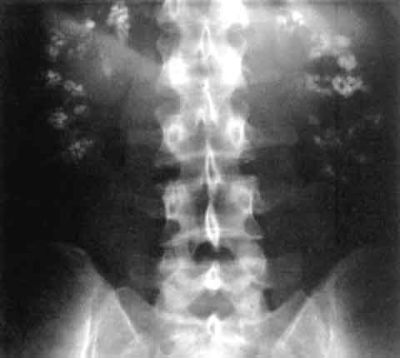
