Uploads by Urology4all
Jump to navigation
Jump to search
This special page shows all uploaded files.
| Date | Name | Thumbnail | Size | Description | Versions |
|---|---|---|---|---|---|
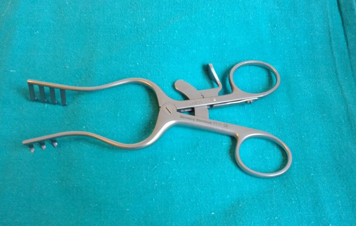
| 18:02, 20 August 2024 | Weitlaner Retractor.jpg (file) |  |
168 KB | 1 | |
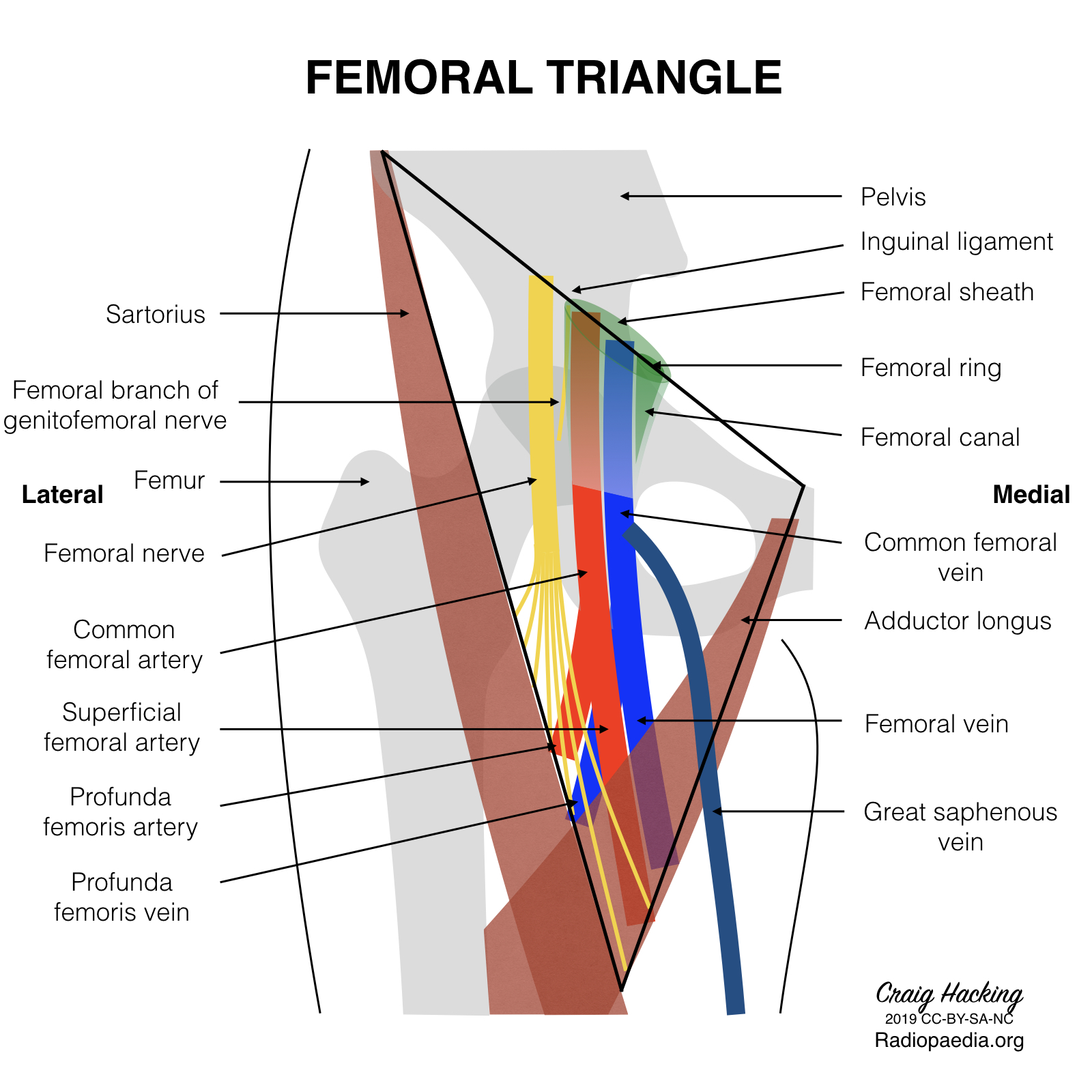
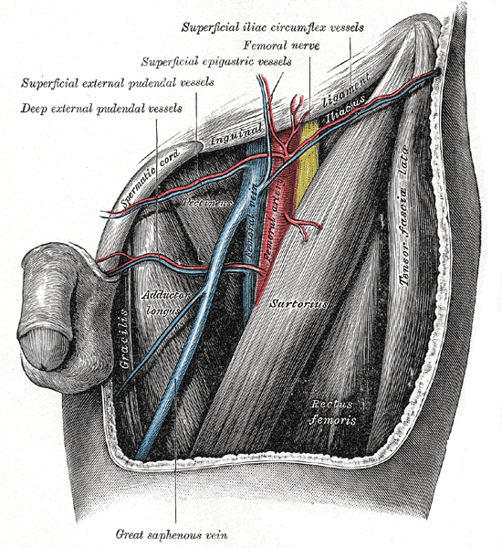
| 11:34, 13 August 2024 | Femoral Triangle Anatomy.jpg (file) |  |
709 KB | 1 | |
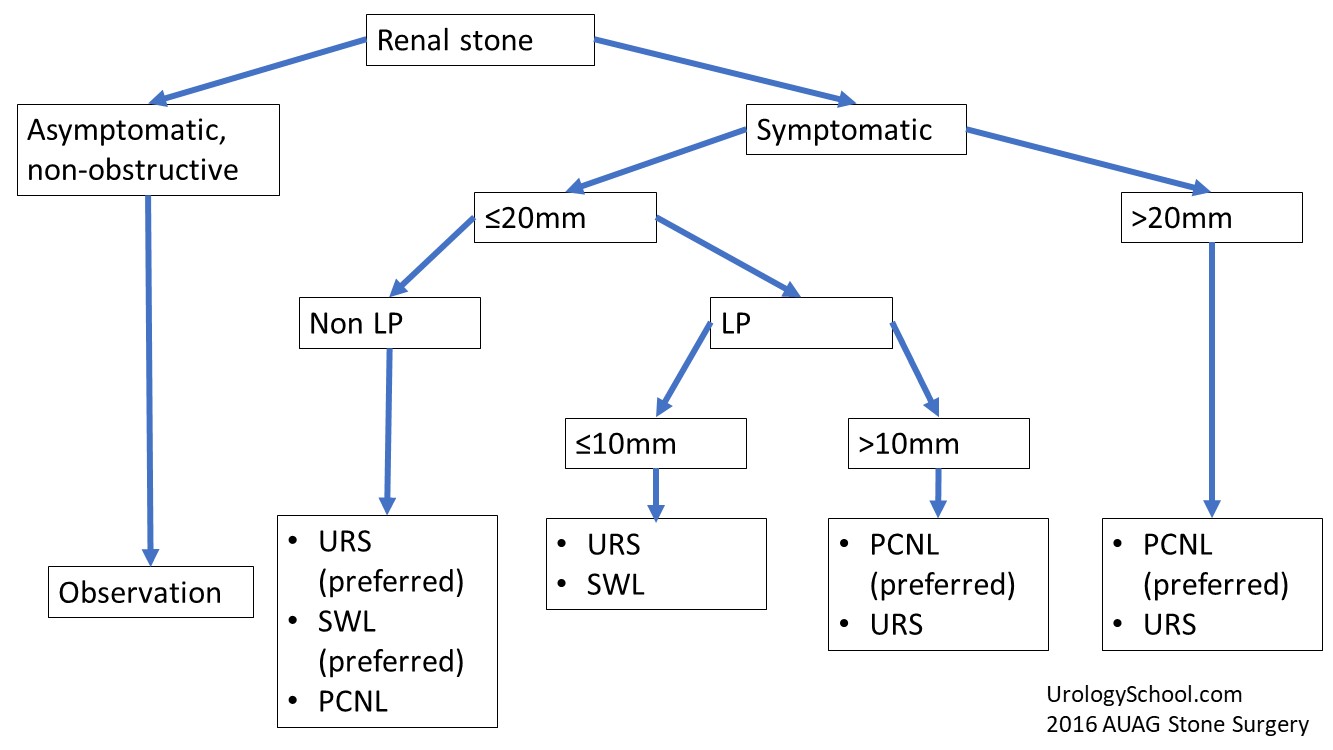
| 13:53, 11 August 2024 | 2019auastonesxpathway.jpg (file) |  |
104 KB | 1 | |
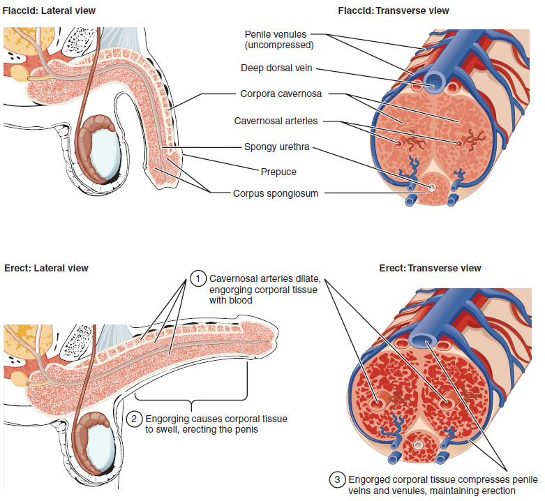
| 19:08, 20 March 2024 | Figure 28 01 06.jpg (file) |  |
295 KB | 1 | |
| 18:24, 20 March 2024 | 43414588252 df2480a453 o.jpg (file) |  |
148 KB | 1 | |
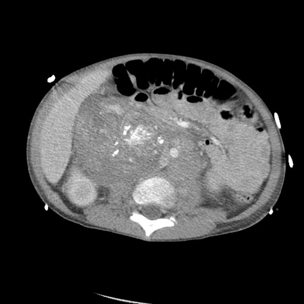
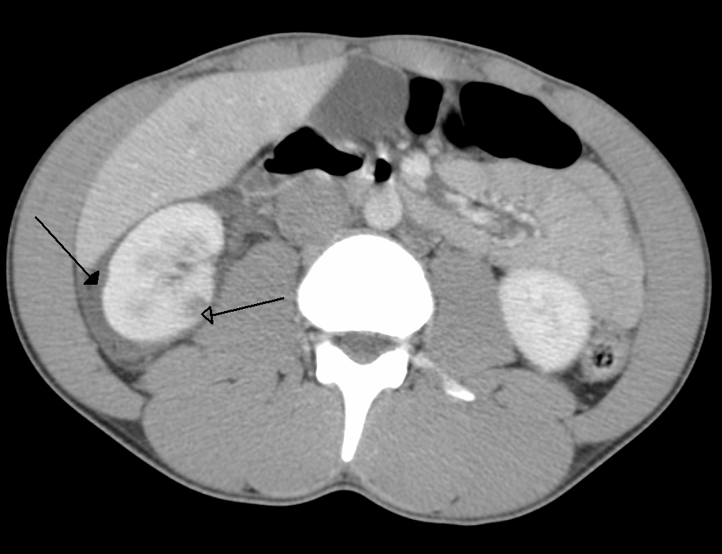
| 17:20, 20 March 2024 | Neuroblastoma 103.jpg (file) |  |
51 KB | 1 | |
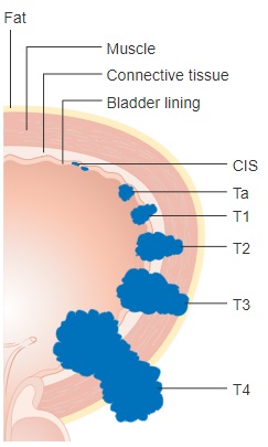
| 18:59, 17 March 2024 | Diagram showing the T stages of bladder cancer CRUK 372.jpg (file) |  |
25 KB | 1 | |
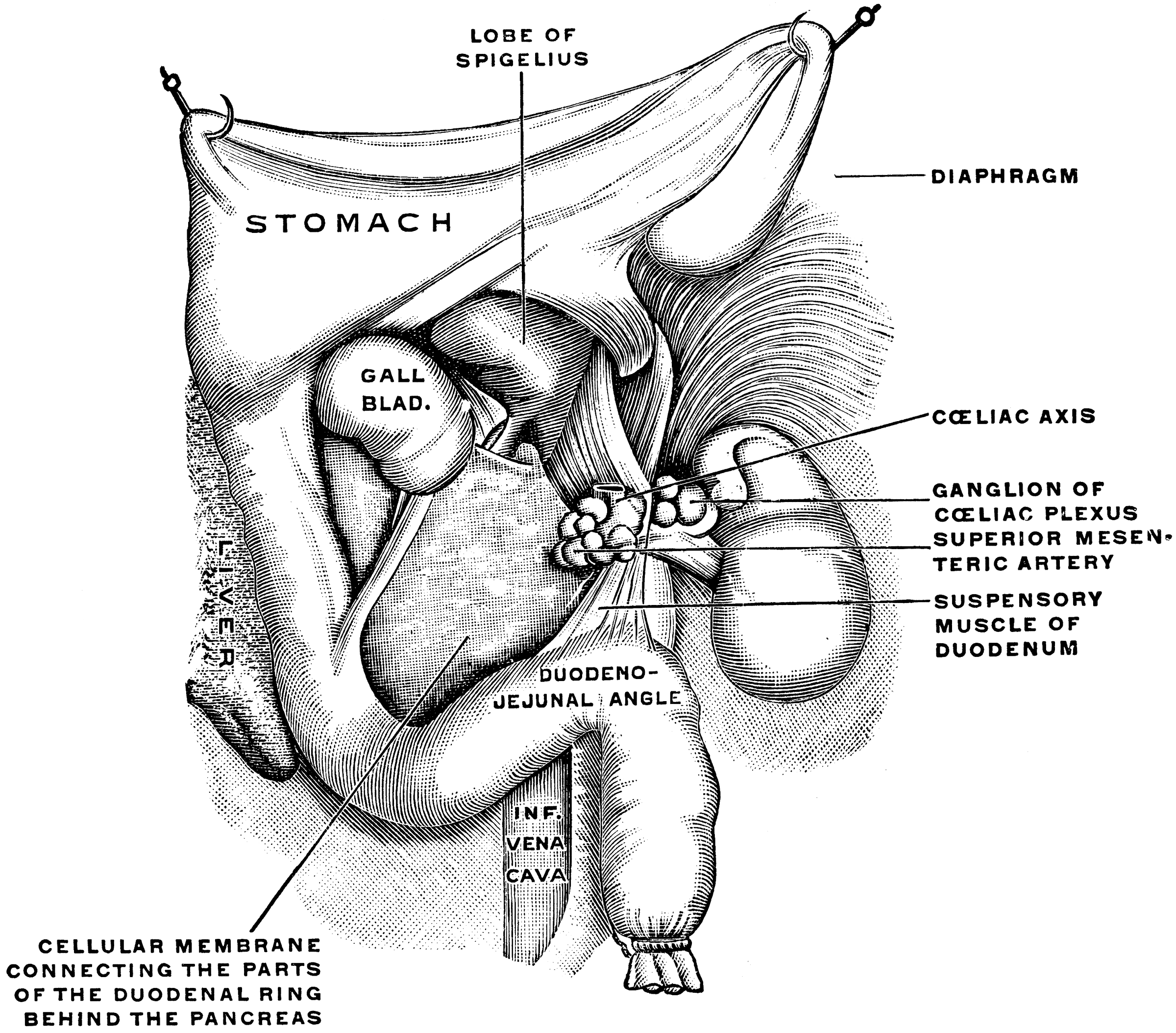
| 10:36, 16 March 2024 | Gray 1913 1285.png (file) |  |
4.83 MB | 1 | |
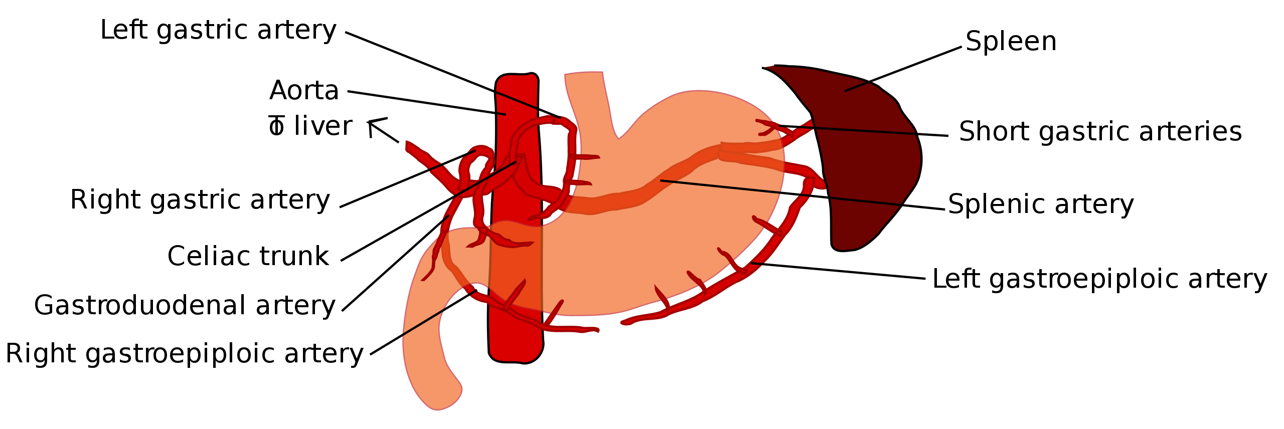
| 10:32, 16 March 2024 | 2560px-Stomach blood supply.svg.png (file) |  |
192 KB | 1 | |
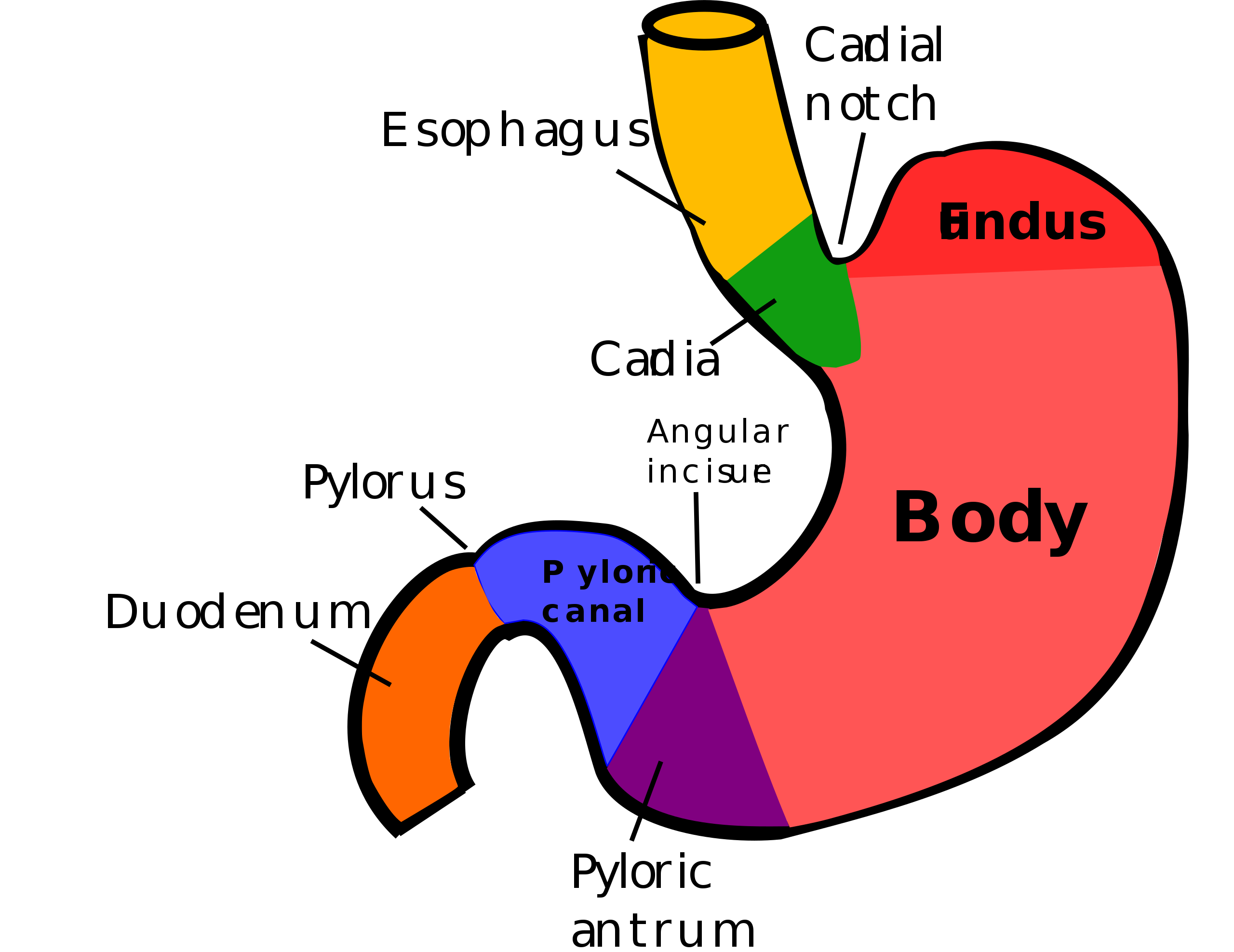
| 10:30, 16 March 2024 | 2560px-Regions of stomach.svg.png (file) |  |
197 KB | 1 | |
| 09:51, 16 March 2024 | Bladder Diverticulum.jpg (file) |  |
379 KB | 1 | |
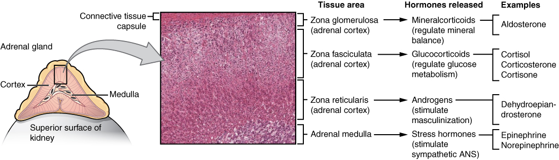
| 05:25, 16 March 2024 | 1818 The Adrenal Glands.jpg (file) |  |
250 KB | 1 | |
| 05:05, 16 March 2024 | TAJU A 1589748 F0005 OC.jpg (file) |  |
27 KB | 1 | |
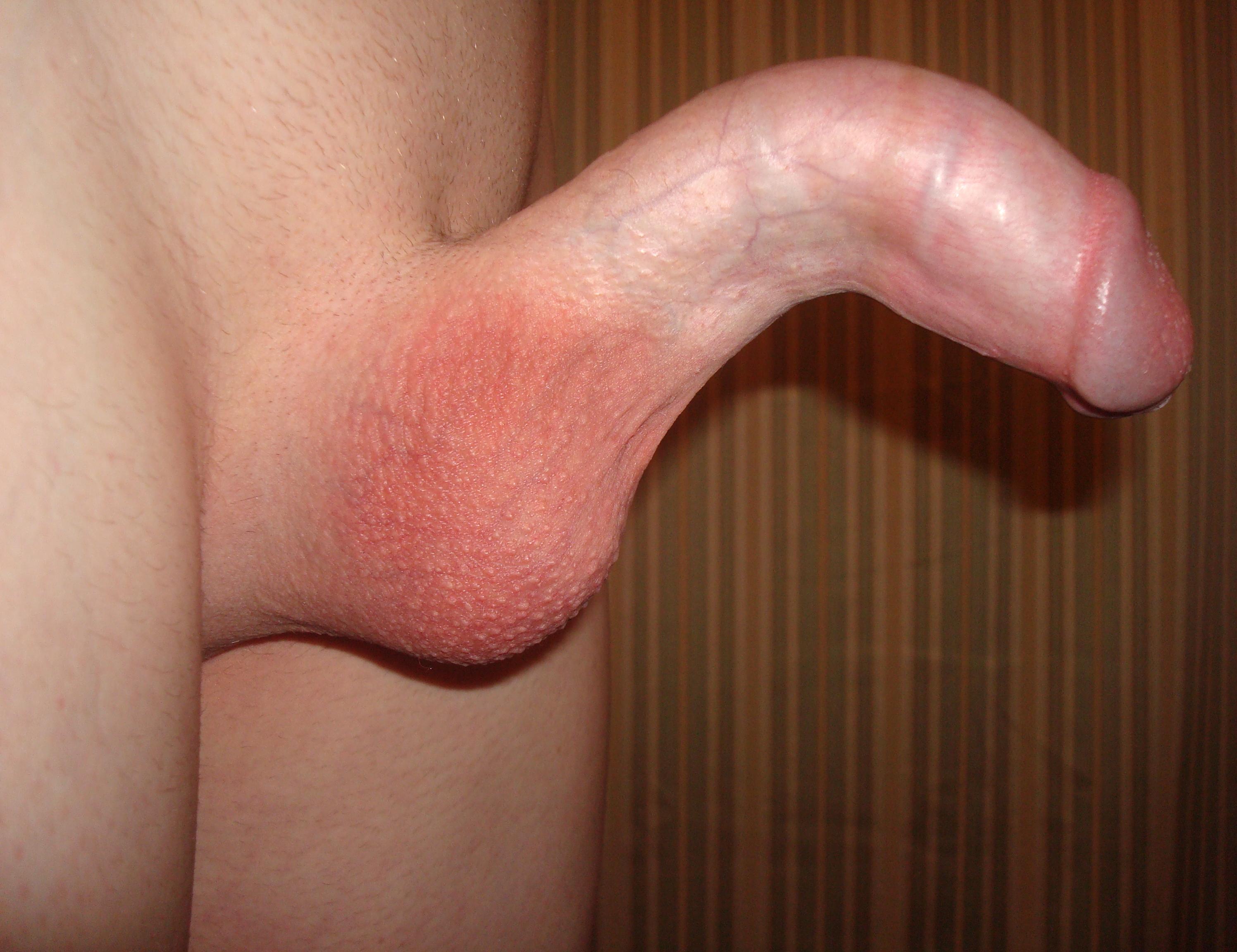
| 10:43, 14 March 2024 | Peyronie's disease.jpg (file) |  |
2.27 MB | 1 | |
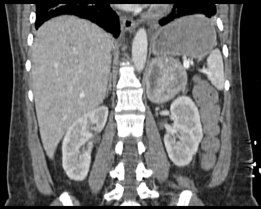
| 17:17, 13 March 2024 | Phaeochromozytoma CT coronal.jpg (file) |  |
107 KB | 1 | |
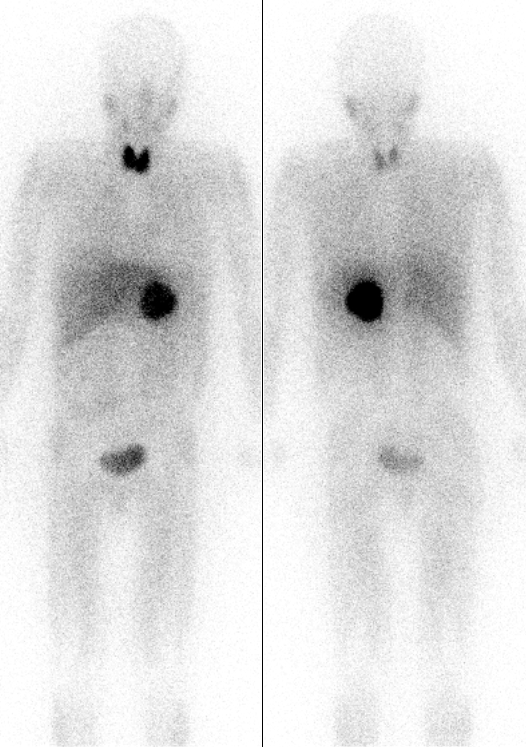
| 17:14, 13 March 2024 | Pheochromocytoma Scan.jpg (file) |  |
244 KB | 1 | |
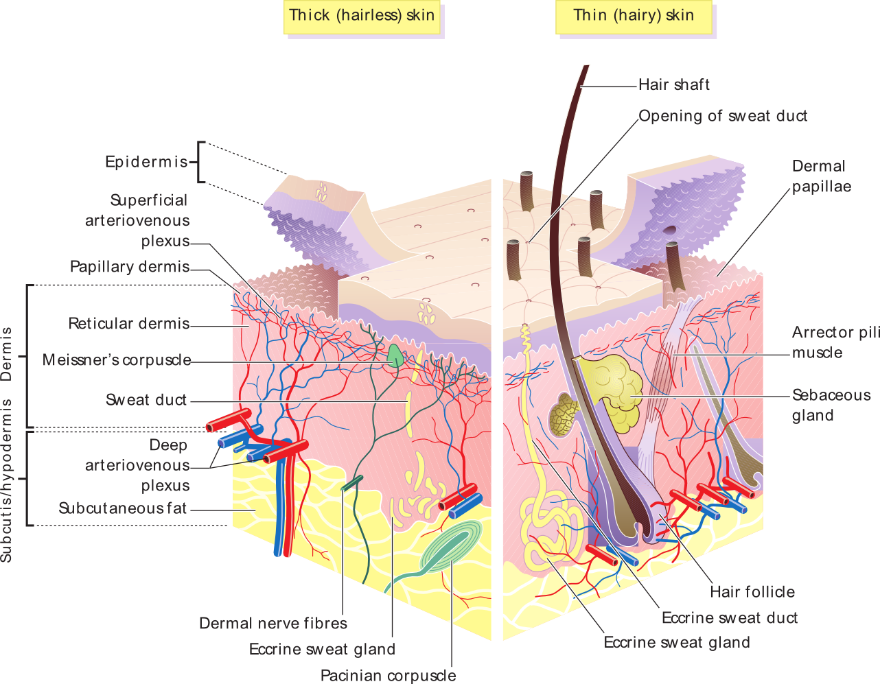
| 08:36, 13 March 2024 | Skin layers.png (file) |  |
621 KB | 1 | |
| 08:39, 12 March 2024 | Stellate scar in right renal mass.jpg (file) |  |
69 KB | 1 | |
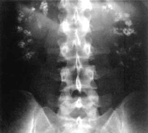
| 12:42, 11 March 2024 | Nephrocalcinosis.jpg (file) |  |
9 KB | 1 | |
| 19:53, 20 February 2024 | CT colovesical fistila.jpg (file) |  |
122 KB | 1 | |
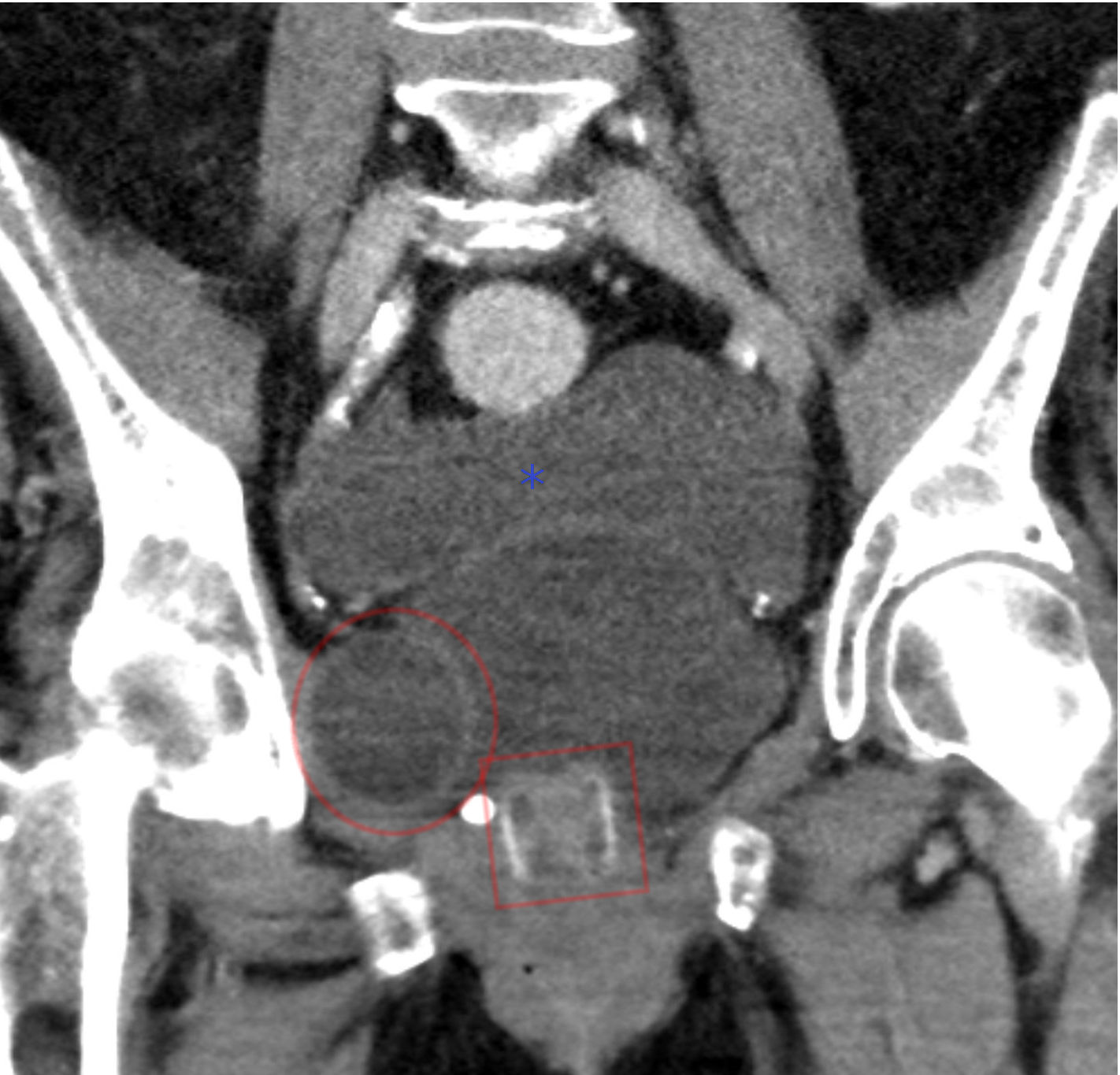
| 19:54, 13 February 2024 | Artificial urethral sphincter - CT coronar 001.jpg (file) |  |
178 KB | 1 | |
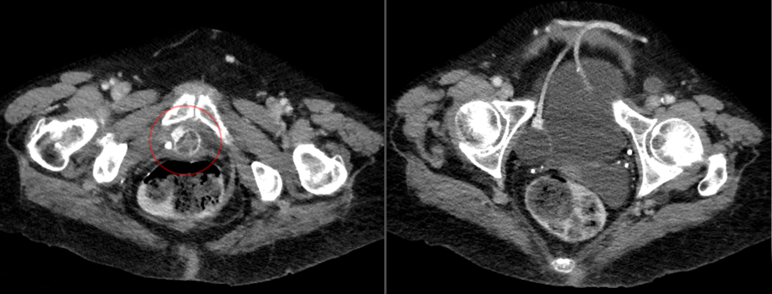
| 19:53, 13 February 2024 | Artificial urethral sphincter - CT axial 001.jpg (file) |  |
307 KB | 1 | |
| 19:50, 13 February 2024 | Blausen 0059 ArtificialUrinarySphincter.png (file) |  |
471 KB | 1 | |
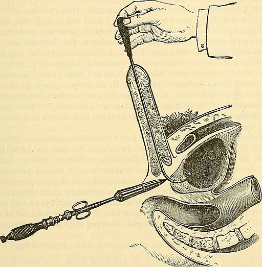
| 20:25, 30 January 2024 | Dolbeau technique of perineal lithotomy.jpg (file) |  |
82 KB | 1 | |
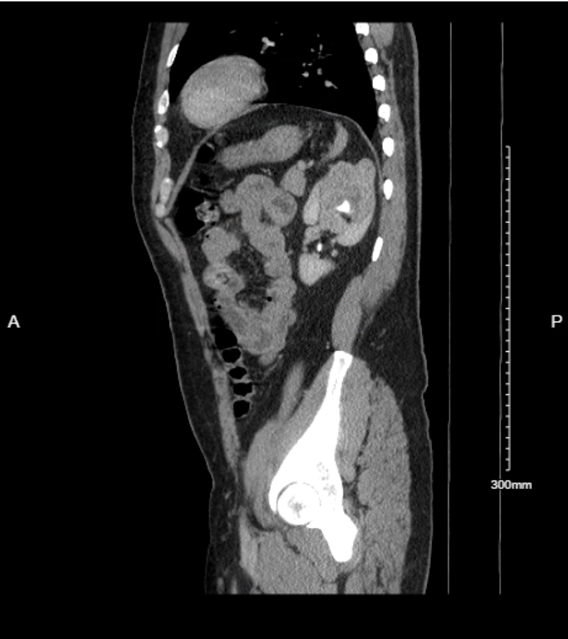
| 20:51, 6 January 2024 | Kidney CT Mass Sagittal.png (file) |  |
312 KB | 1 | |
| 20:43, 6 January 2024 | Kidney CT Mass Axial.png (file) |  |
287 KB | 1 | |
| 14:34, 16 August 2023 | Wiki Penis Base Cross Section.jpg (file) |  |
185 KB | 1 | |
| 14:21, 16 August 2023 | Penis Anatomy Cross Section.png (file) |  |
69 KB | 1 | |
| 12:48, 30 January 2023 | Prostate zones.png (file) |  |
207 KB | 1 | |
| 13:08, 9 November 2022 | Femoral triangle.png (file) |  |
134 KB | 1 | |
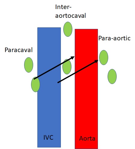
| 18:00, 12 October 2022 | Retroperitoneal lymph flow.jpg (file) |  |
28 KB | 1 | |
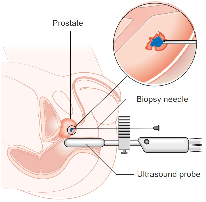
| 17:03, 10 October 2022 | Diagram showing a transperineal prostate biopsy CRUK 473.jpg (file) | 114 KB | 1 | ||
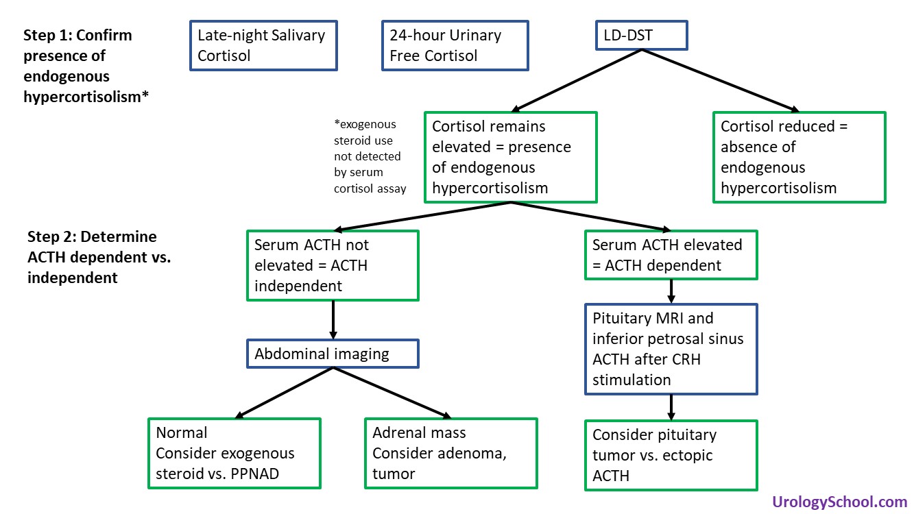
| 11:28, 29 September 2022 | Hypercortisolism.jpg (file) |  |
153 KB | 1 | |
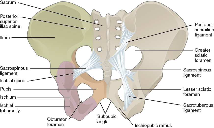
| 11:27, 21 September 2022 | 817 Ligaments of Pelvis.jpg (file) |  |
222 KB | 1 | |
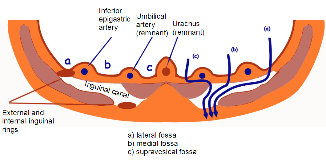
| 11:09, 21 September 2022 | Inguinal fossae.png (file) |  |
111 KB | 1 | |
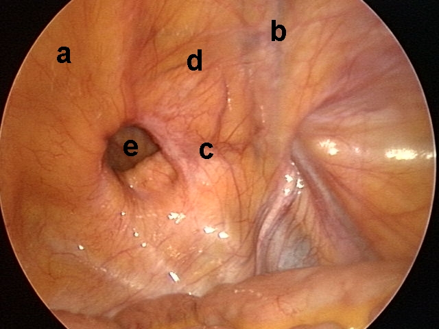
| 11:06, 21 September 2022 | Laparoscopic view of inguinal region.jpg (file) |  |
155 KB | 1 | |
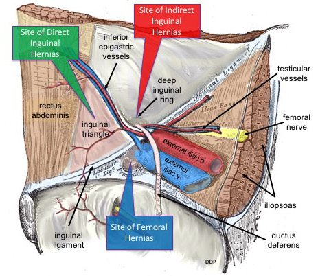
| 11:05, 21 September 2022 | Common Sites of Lower Abdominal Hernias.jpg (file) |  |
88 KB | 1 | |
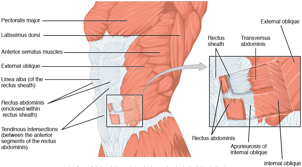
| 11:01, 21 September 2022 | 1112 Muscles of the Abdomen Anterolateral.png (file) |  |
450 KB | 1 | |
| 14:52, 10 December 2021 | Abdotrauma.png (file) |  |
434 KB | 1 | |
| 14:48, 10 December 2021 | Leftrenalarteryinjury.png (file) |  |
834 KB | 1 | |
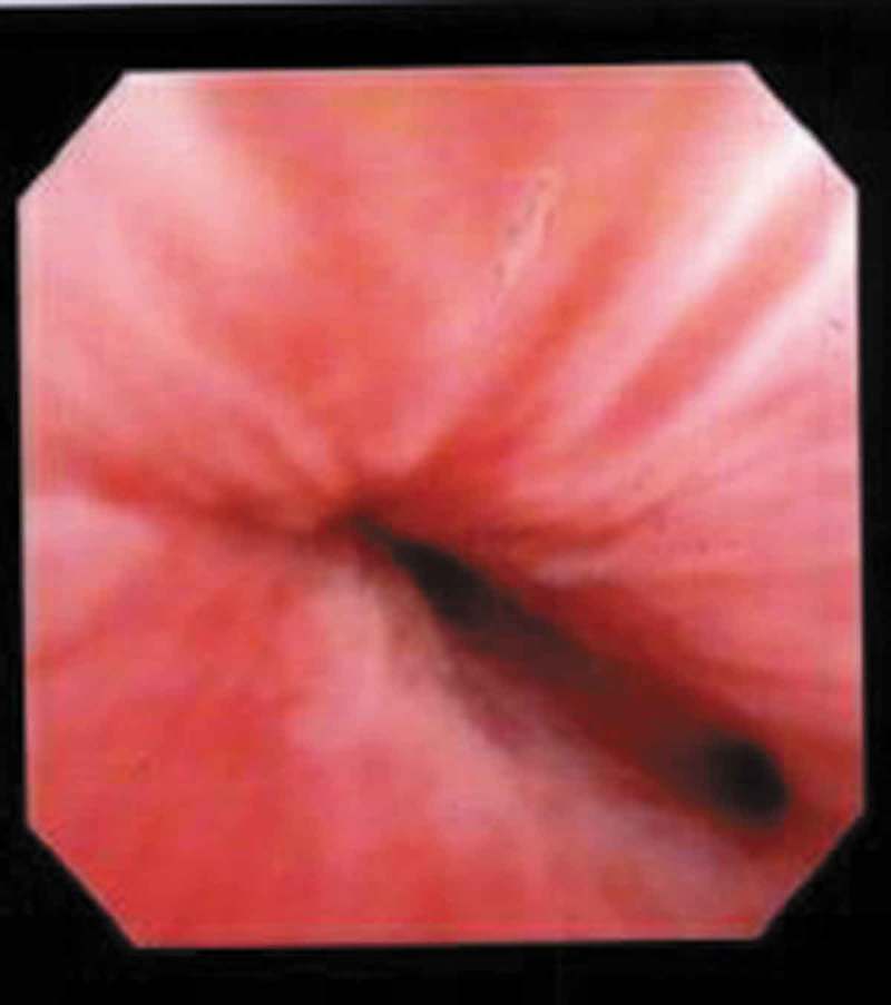
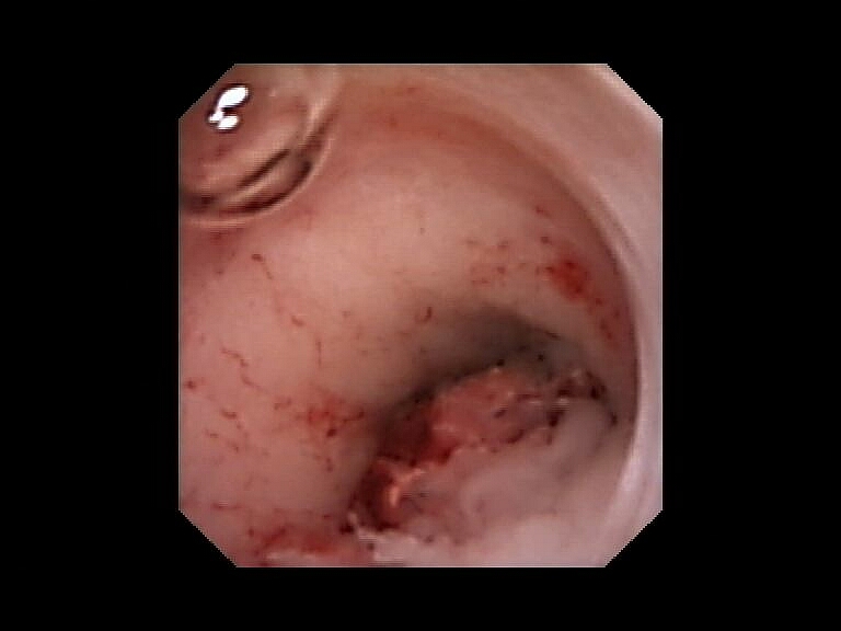
| 12:52, 10 December 2021 | Cystoscopy - Uretereal Cancer.jpg (file) |  |
105 KB | 1 | |
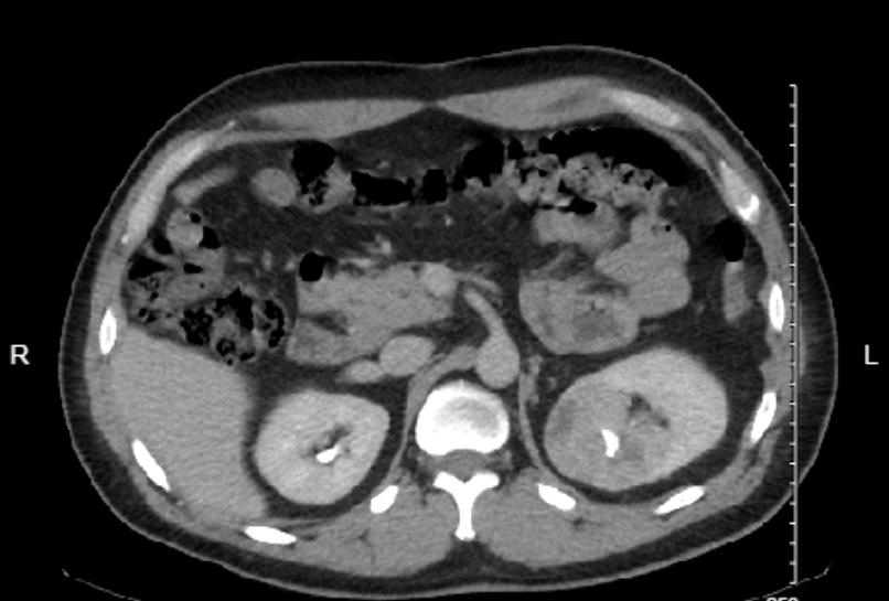
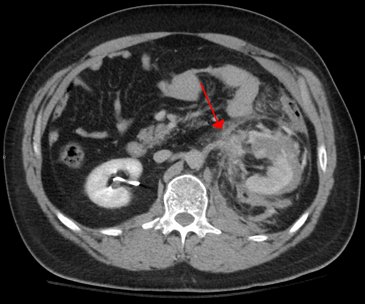
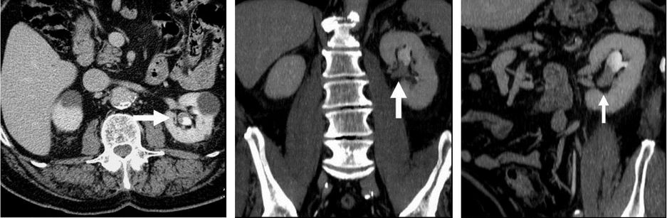
| 12:46, 10 December 2021 | Renal parenchymal phase CT of transitional cell carcinoma.jpg (file) | 99 KB | Description English: Renal parenchymal phase CT of transitional cell carcinoma. For context, see en:Computed tomography of the abdomen and pelvis. Date 15 May 2013 Source Dongqing Wang (2013) Selected Topics on Computed Tomography ISBN: 9789535111023. License: CC-BY-3.0. Chapter 1: "Computed Tomography in Abdominal Imaging: How to Gain Maximum Diagnostic Information at the Lowest Radiation Dose" by Kristie Guite, Louis Hinshaw and Fred Lee. DOI: 10.5772/55903 Author Kristie Guite, Louis Hins... | 1 |