ANATOMY: TESTICLE
Gross structure
- Normal
- Dimensions: length 4-5 cm x width 3 cm x depth 2.5 cm
- Volume: 15-25 mL
- Although ethnic and racial differences do exist, a normal testis volume is generally above 16 mL and averages 20 mL.
- Right testicle hangs lower compared to the left in ≈85% of men
- Appendix testis
- A small pedunculated or sessile body at the upper pole of the testis
- Mullerian (paramesonephric) duct remnant; recall that appendix epididymis is a Wolffian (mesonephric) duct remant
- The epididymis attaches on the posterolateral aspect of the testis


Microanatomic architecture
- Comprised of (2):
- Seminiferous tubules
- Interstitial tissue
- Seminiferous tubules
(seminal vesicle lobules in above diagram)
- Contain Spermatogenic cells and supporting cells, including Sertoli cells
- Spermatogenic cells
- Comprise the bulk of the testicular volume
- Sertoli cells
- Rest on the tubular basement membrane
- Support spermatogenesis
- Secrete Inhibin which inhibits FSH secretion
- Tight junctions between Sertoli cells provide an intracellular barrier, known as the blood-testis barrier, that allows for spermatogenesis to occur in an immune privileged site
- This forms a high-resistance barrier that prevents the deep penetration of most drugs, molecules, and electron-opaque tracers into the seminiferous epithelium from the testicular interstitium.
- Sertoli cell tight junctions also segregate pre-meiotic germ cells (spermatogonia) from meiotic and post-meiotic germ cells.
- The blood-testis barrier develops during puberty
- Clinical implication: since post-meiotic germ cells only exist after puberty, testicular insult (biopsy, torsion, trauma) before puberty will not induce anti-sperm antibodies
- Spermatogenic cells
- The convoluted seminiferous tubules become straight at their apices and then enter the mediastinum testis to form an anastomosing network of tubules, called the rete testis. The rete testis form 12-20 efferent ductules that anastamose with in head of the epididymis
- Structures the sperm passes through in sequence: seminiferous tubules-->straight tubules (tubuli recti)-->rete testis-->efferent ductules-->head, body, tail of epididymis-->vas deferens-->ejaculatory duct-->prostatic urethra
- Contain Spermatogenic cells and supporting cells, including Sertoli cells

Animation showing the flow of spermatozoa through the system of ducts and tubules.
A.) Blood vessels; B.) Head of epididymis; C.) Efferent ductules; D.) Seminiferous tubules; E.) Parietal lamina of tunica vaginalis; F.) Visceral lamina of tunica vaginalis; G.) Cavity of tunica vaginalis; H.) Tunica albuginea; I.) Lobule of testis; J.) Tail of epididymis; K.) Body of epididymis; L.) Mediastinum; M.) Vas deferens
Source: Wikipedia
- Interstitial tissue
- Around the seminiferous tubules
- Makes up to 20-30% of the testicular volume
- Contains Leydig cells, mast cells, macrophages, nerves, blood vessels, and lymphatic vessels
- Leydig cells
- Produce testosterone and activin
- Activin stimulates production of FSH
- Produce testosterone and activin
Vasculature
- Arterial supply (3):
- Testicular (internal spermatic) artery
- Arises from the abdominal aorta
- Crosses psoas muscle and the ureter to reach the internal inguinal ring to enter the spermatic cord
- Artery of the vas deferens (vasal/deferential artery)
- Arises from the superior vesical artery (usually, but can arise from the inferior vesical artery), which arises from the internal iliac artery
- Cremasteric (external spermatic) artery
- Arises from the inferior epigastric artery (Cream in the Belly), which arises from the external iliac artery
- Testicular (internal spermatic) artery
Testicular artery
Main blood supply to the testicle
- Diameter of the testicular artery > (diameter of artery of vas + cremasteric arteries)
- In case of testicular artery ligation, the deferential and cremasteric arteries can potentially provide adequate blood supply to the testis; however, atrophy and/or azoospermia has resulted from testicular artery ligation
- Because the deferential artery may have been compromised during a prior vasectomy, careful attention is needed to preserve the testicular artery in future surgeries, such as varicocelectomy, in men with a prior vasectomy
- Penetrates the tunica albuginea and then travel inferiorly along the posterior surface of the testis within the parenchyma. Branching arteries pass anteriorly over the testicular parenchyma. Major testicular artery branches also travel over the inferior pole of the testis, pass anteriorly, and branch out over the surface of the testis.
- Clinical implication: testicular biopsy should target the midsection of the testis because this area has relatively fewer vessels compared with superior or inferior areas
- Supplies the tunica testis vasculosa in the anterior portion of the upper pole of the testis and the anterior, medial, and lateral portions of the lower pole of the testis
- Genital branch of the genitofemoral nerve travels along the testicular artery to reach the testis
- Primarily supplies sensation to the parietal and visceral tunica vaginalis and the overlying scrotum
- Venous drainage
- Unlike most other venous patterns in the body, veins within the testis do not travel with their corresponding arteries.
- Small parenchymal veins either drain into a group of veins near the mediastinum testis, or they drain into veins on the surface of the testis. These two groups of veins anastomose with each other and the deferential veins to form the pampiniform plexus
- Pampiniform plexus
- A network of testicular veins that anastomose as they ascend surrounding the testicular artery
- Allows for a counter-current heat exchange that cools the blood flow within the testicular artery and keeps the testis 2-4°C lower than rectal temperature
- Ultimately, these veins join one another to form 2-3 veins at the level of the inguinal canal, and then they form one vein (gonadal vein) that ascends to drain into the inferior vena cava on the right and into the renal vein on the left side
- Dilated in a varicocele (see Varicocelectomy Chapter Notes); there may be variations where the testicular veins can anastomose to other veins and this can result in recurrence of varicocele following ablation.
- Deferential veins
- Follow the vas deferens and empty into the internal iliac (hypogastric) veins
- Spared during varicocele ligation surgery
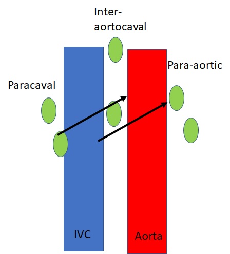
Lymphatic supply
- Lymphatic channels ascend within the spermatic cord after leaving the testis
- Drain into (3):
- Para-aortic lymph nodes
- Interaortocaval lymph nodes
- Para-caval lymph nodes
- Right to left direction of flow
- Right testicle drains predominantly to inter-aortocaval nodes and secondarily to para-aortic and para-caval nodes
- Left testicle drains predominantl to para-aortic nodes and secondarily to inter-aortocaval nodes
Innervation
- No somatic innervation of the testicle
- Autonomic innervation to the testis and epididymis
- Arises in the renal and aortic plexuses and travels alongside the gonadal vessels
- Nerves contributing to chronic orchialgia (3):
- Peri-vasal complex
- Intracremasteric complex
- Posterior peri-arterial/lipomatous complex
Radiology
- Ultrasound
- Scrotal US uses high-frequency transducers (7.5 to 10 MHz)
- An anechoic area between the echogenic scrotal wall and testicle is commonly visualized, which represents a small amount of physiologic fluid between the visceral and parietal layers of the tunica vaginalis
Questions
- What is the normal volume of the testicle?
- Which cells are responsible for the blood-testis barrier?
- When does the blood-testis barrier develop?
- What is the arterial blood supply to the testicle and what is the origin of each artery?
- Where should a biopsy of the testicle be done to reduce the risk of bleeding?
- Where do the testicular lymphatics drain?
- What are the nerves contributing to chronic orchalgia?
Answers
- What is the normal volume of the testicle?
- 15-25cc
- Which cells are responsible for the blood-testis barrier?
- Sertoli cells
- When does the blood-testis barrier develop?
- Puberty
- What is the arterial blood supply to the testicle and what is the origin of each artery?
- Testicular artery from aorta
- Deferential artery from superior vesical artery
- Cremesteric artery from inferior epigastric
- Where should a biopsy of the testicle be done to reduce the risk of bleeding?
- Middle of testicle
- Where do the testicular lymphatics drain?
- Drain into the para-aortic, interaortocaval, and para-caval lymph nodes
- Right to left direction of flow
- Right testicle drains predominantly to inter-aortocaval nodes and secondarily to para-aortic and para-caval nodes
- Left testicle drains predominantl to para-aortic nodes and secondarily to inter-aortocaval nodes
- What are the nerves contributing to chronic orchalgia?
- Perivasal complex
- Intracremasteric complex
- Posterior periarterial/lipomatous complex
References
- Wein AJ, Kavoussi LR, Partin AW, Peters CA (eds): CAMPBELL-WALSH UROLOGY, ed 11. Philadelphia, Elsevier, 2015, chap 21