In cases of VHL the risk of malignancy is low but pheochromocytoma is characterized as producing norepinephrine.
Unlike pheochromocytomas in patients with VHL, MEN2 and NF1 predominantly produce epinephrine.
Compared to sporadic cases, in familial syndromes, the pheochromocytomas are almost always bilateral and more frequently malignant. Clinical manifestations are similar.
Can have heterogenous clinical behavior due to the variability in the amount and ratio of the different catecholamines (norepinephrine, epinephrine, dopamine)secreted:
Norepinephrine-predominant tumours (e.g., patients with von Hippel- Lindau [VHL] syndrome) have hypertension and sweating because of norepinephrine's vasoconstricting action through the α adrenoreceptor
Epinephrine-predominant tumours (rare, usually limited to adrenals or the organ of Zuckerkandl)have syncope or hypotensive episodes because of epinephrine’s vasodilatory action through the β2 receptor
Agonist potency order:
α1: epinephrine ≥ norepinephrine >> isoprenaline
α2: epinephrine ≥ norepinephrine >> isoprenaline
β1: isoprenaline > epinephrine = norepinephrine
β2: isoprenaline > epinephrine >> norepinephrine
β3: isoprenaline = norepinephrine > epinephrine
Episodic hypertensive episodes may be triggered by events such as induction of anesthesia, labor and delivery, instrumentation and biopsy of the tumor, strenuous physical activity and consumption of tyramine rich foods, such as red wine, chocolate and cheeses.
Another serious clinical presentation may result from catecholamine induced cardiomyopathy when patients present with congestive heart failure and cardiac arrhythmias.
The enzyme phenylethanolamine-N-methyltransferase (PNMT), catalyzes the conversion of norepinephrine to epinephrine, is relatively unique to the adrenal medulla (the brain and organ of Zuckerkandl also express this enzyme).
Localization of PNMT to the adrenal medulla explains why the gland is the primary source of systemic epinephrine, despite the presence of similar chromaffin cells elsewhere in the sympathetic nervous system
Catecholamines (dopamine, norepinephrine, and epinephrine) are produced by pheochromocytomas in varying amounts; release of these compounds into the bloodstream is often paroxysmal
Metanephrines are the methylated metabolites of catecholamines
O-methylation of catecholamines is catalyzed by the COMT enzyme
O-methylation of norepinephrine produces normetanephrine, whereas epinephrine’s methylation results in formation of metanephrine.
Together, normetanephrine and metanephrine are known as metanephrines.
The conversion of catecholamines to metanephrines within pheochromocytomas is an uninterepted process. As such, measurement of plasma concentration of metanephrines is much more sensitive for detecting pheochromocytomas than the measurement of rises in plasma catecholamines, which may be paroxysmal.
In the past, measurement of both urinary and serum catecholamine levels was the mainstay for evaluation of pheochromocytoma. However, these tests had moderated sensitivity and specificity and have been largely replaced by measurements of levels of metanephrines.
Measurement of urinary catecholamines, nevertheless, is still recommended in conjunction with urinary fractionated (see below) metanephrine testing
Measurement of plasma fractionated metanephrines and 24-hour urinary fractionated metanephrines and catecholamines are the mainstay biochemical tests to diagnose pheochromocytoma.
Plasma fractionated metanephrines is used primary in those with high index of suspicion with 24-hour urinary fractionated metanephrines for those with low index.
2011 CUA Incidental Adrenal Mass guidelines recommend screening with a 24-hour urinary fractionated metanephrines and/or catecholamines.
2014 [most recent as of October 2019] Endocrine Society Pheochromocytoma Guidelines recommend plasma free metanephrines or 24-hour urinary fractionated metanephrines
2019 AUA Update on Pheochromocytoma recommended initial screening with plasma free metanephrines. Urinary fractionated metanephrines are an alternative, with slightly lower diagnosis sensitivity.
The term fractionated is used when the laboratory report details not only the amount of each compound type (e.g., metanephrines), but also the relative concentrations of each compound (e.g., normetanephrine and metanephrine).
The blood sample should be drawn after placing an intravenous cannula, dimming the room lights and having the patient lay supine for 30 minutes after minimizing any pain or anxiety.
Prior to the blood draw, patients should be counseled on avoiding caffeine for a minimum of 24 hours
Acetaminophen can produce a false-positive result owing to cross reactivity in the [serum?] assay and should be stopped for at least 5 days before testing.
Tricyclic antidepressants and phenoxybenzamine should also be stopped, because these have been shown to be responsible for false-positive results.
Usual antihypertensive therapy can be continued.
Although β-blockade can potentially result in a false-positive test result, the current recommendation is to stop the medication only on repeat testing
Vanillylmandelic Acid (VMA) Testing
VMA is the primary end metabolite of catecholamines
The sympathetic nervous system lacks the ability to produce epinephrine (lacks PNMT enzyme) and therefore contributes to the serum level of only normetanephrine (from norepinephrine) but not metanephrine (from epinephrine). Indeed, > 90% of metanephrine (an epinephrine metabolite) and some 20% or more of normetanephrine (a norepinephrine metabolite) in the bloodstream are derived from the adrenal medulla (PNMT is present in the brain and organ of Zuckerkandl). Therefore the relative rise of VMA levels, the combined total end metabolite of norepienphrine and epinephrine, in the presence of a pheochromocytoma is much less dramatic than the rise seen in the levels of metanephrines (from epinephrine), and therefore, the sensitivity of urine VMA levels is low. However, the specificity of VMA is high, especially in nonfamilial cases.
Oral clonidine testing
Used to distinguish suspected pheochromocytoma vs. essential hypertension in patients with minimally elevated plasma catecholamines.
Patients with suspected pheochromocytoma usually present with elevated plasma catecholamines. However, they can rarely present with normal or mildly elevated plasma catecholamines.
The oral clonidine test can help distinguish whether the signs and symptoms and minimally elevated plasma catecholamines are related to pheochromocytoma vs. essential hypertension, oral clonidine test.
Patients with essential hypertension will experience a significant drop in norepinephrine due to suppression of production by the sympathetic nervous system, while those with pheochromocytoma will not.
18F-FDG PET (fluorine-18 fluorodeoxyglucose positron emission tomography)
Gold standard imaging modality for definitive staging in patients with pheochromocytoma.
Superior test characteristics to CT, MRI, and metaiodobenzylguanidine (MIBG) scintigraphy.
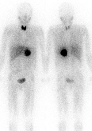
Better accuracy than 123I-MIBG in nearly all patients, especially for identification of metastatic disease.Pheochromocytoma (dark circular shadow near body center) localized by MIBG scintigraphy. See corresponding CT below. Source: Wikipedia
MIBG
Utilizes a small-molecule analog of norepinephrine
High specificity but low sensitivity for diagnostic disease identification.
Useful modality when a suspected pheochromocytoma cannot be localized or when metastatic disease is suspected.
In the most common and urologically most relevant clinical scenario, a solitary adrenal mass on cross sectional imaging in the setting of a biochemical evaluation indicative of pheochromocytoma, MIBG or 18F-FDG PET may be safely be omittedbecause these functional studies only serve to confirm what is already known and do not alter management.However, MIBG or 18F-FDG PET imaging for large (>5 cm) tumors is likely prudent to assess for metastatic disease before surgery and thereby counsel the patient appropriately
MRI
Distinct low signal intensity on T1-weighted imaging
High signal intensity on T2-weighted imaging.
CT
On unenhanced CT, pheochromocytomas are typically > 10 HU (mean ≈35 HU) given their rich vascularity and low lipid content. This can help differentiate them from lipid-rich adenomas
If the lesion is not an adenoma, an adrenal mass protocol CT with IV contrast allows for evaluation of tumor washout.
Benign adrenal lesions wash out >50% on delayed imaging while pheochromocytoma, adrenocortical carcinoma and metastatic tumors do not.
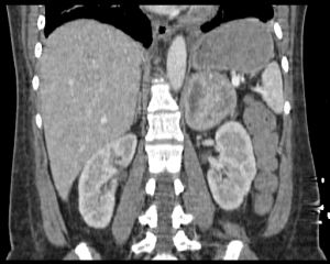
CT scan with enhancement demonstrating suspicous left adrenal mass. See corresponding MIBG scan above. Source: WikipediaPheochromocytomas usually measure greater than 10 HU on unenhanced CT and >100 HU on contrast imaging, and are often well circumscribed in appearance with or without necrotic or cystic elements. Nevertheless, any evaluation of an adenoma should still include testing for plasma free metanephrines to rule out pheochromocytoma.
CT scan with enhancement demonstrating suspicious left adrenal mass. See corresponding MIBG scan above. Source: Wikipedia
Investigation for familial syndromes is warranted in a patient younger than 50 years with a significant family history of an extraadrenal pheochromocytoma (hereditary paraganglioma syndrome), or bilateral or multifocal tumors.
If a mutation is identified, screening should also be offered to asymptomatic at risk family members.
Pre-operative cardiology or anesthesia consultation because of risk for cardiomyopathy
Preoperative cardiac workup, including electrocardiography and echocardiography, and assessment of hypertension-induced end-organ dysfunction are indicated.
Restoration intravascular volume
Pre-operative medications (α-Blockade followed by β-blockade)
All patients with pheochromocytoma and an abnormal metabolic evaluation undergo preoperative catecholamine blockade, including patients who do not exhibit evidence of blood pressure elevation and lack classic symptomatology.
Catecholamine release during intraoperative tumor manipulation can result in hazardous blood pressure elevation and cardiac arrhythmias.
Recent data suggest that preoperative α-blockade may not be necessary in normotensive asymptomatic patients
α-Blockade
Helps in both hemodynamic and glucose control
Phenoxybenzamine
Most common α- blocker used for preoperative catecholamine blockade of pheochromocytoma.
Intraoperative catecholamine surges typically do not override its actions, because reversal of the blockade is possible only through synthesis of new receptor molecules.
Non-selective nature may lead to tachycardia and β-adrenergic blockade may be necessary
Prolonged hypotension in the immediate postoperative period and central nervous system effects such as somnolence may be expected
Newer selective and competitive α1-adrenergic blockers such as doxazosin, prazosin, and terazosin obviate the drug-induced need for β-blockade.
Started 7-14 days before surgery.
Can be started at 10 mg twice daily with a stepwise increase of 10 to 20 mg every 2 to 3 days until a final dose of 1 mg/kg if tolerated.
During this time blood pressure checks should be conducted at least 3 times a day.
The last dose of phenoxybenzamine is usually given on the night before surgery, and the next morning’s dose is withheld to minimize potentially prolonged hypotension after tumor resection.
If phenoxybenzamine not effective for blockade, start metyrosine
MOA of metyrosine: blocks the biosynthesis of catecholamines by inhibiting the conversion of tyrosine to L-dopa
Generally added for extensive disease with large increases in catecholamines.
Acute hypertensive attacks can also be treated with a short-acting alpha blocker such as phentolamine.
β-blockade
Must be given with caution in patients with myocardial depression
Should never be started before appropriate α-blockade
In the absence of α-blockade, β antagonists cause a potentiation of the action of epinephrine on the α1 receptors, resulting in hypertension, owing to blockade of the arteriolar dilation at the β2 receptor. For this reason, selective β1 adrenoreceptor blockers, such as atenolol and metoprolol, are usually preferred.
May be added when (2)
Systolic blood pressure is <100 mmHg
Tachycardia or reflex tachycardia develops.
Calcium channel blockers
Some studies report that sole use of calcium channel blockers is sufficient for safe pheochromocytomas resection. This approach avoids the reflex tachycardia and postoperative hypotension that are seen with use of phenoxybenzamine. This strategy be reserved for patients who are normotensive with paroxysmal hypertension and a normal baseline blood pressure.
Usually, preoperative calcium channel blockade for 2 weeks is sufficient.
In the immediate postoperative period, consider overnight ICU admission for active monitoring
If phenoxybenzamine was used for preoperative α-blockade, hypotension is common, given the lasting effects of the agent. Moreover, in a high catecholamine state, α2-adrenoreceptor stimulation inhibits insulin release. The withdrawal of this adrenergic stimulus after tumor resection may result in rebound hyperinsulinemia and subsequent hypoglycemia
Repeat metabolic testing should be performed ≈2 weeks after adrenalectomy to document normalization of catecholamine levels
Postoperative cross-sectional imaging is reasonable to document tumor resection and appropriate healing of the resection bed.
Subsequent imaging should be guided by results of biochemical testing
Annual biochemical follow-up is mandatory for all patients with resected pheochromocytoma.
Lifelong screening for recurrence is recommended
10-year recurrence rates are as high as 16%.
No consensus on follow-up protocols exists.
In a patient with persistent hypertension 2-3 months after adrenalectomy, residual tumor somewhere else in the body must be considered. The patient should initially have plasma free metanephrine levels measured. If this is abnormal, MIBG scan may be helpful in identifying the location of this lesion.
Treatment of Hereditary Pheochromocytoma[edit | edit source]
Given that for patients with MEN-2 and VHL, the risk of malignancy is low whereas the risk of bilateral disease is significant, partial cortical-sparing adrenalectomy has been advocated. This strategy is used to avoid lifelong hormonal replacement, with its associated morbidity
Currently, malignant pheochromocytoma can only be defined by the presence of clinical metastases.
A number of pathologic criteria to differentiate benign from malignant disease have been proposed, but to date there is no criterion agreed on.
Currently therapy for metastatic pheochromocytoma is largely palliative.
Surgical metastasectomy of resectable disease is the standard of care
Little evidence exists to demonstrate that it prolongs patient survival or is more effective for symptomatic relief than medical treatment with α/β-blockade and α-methyl-p-tyrosine
Chemotherapy is primarily used in patients in whom MIBG therapy has failed or in those whose tumors do not demonstrate MIBG uptake on initial MIBG imaging studies.
Treatment of Pheochromocytoma in Pregnancy[edit | edit source]
In a late term pregnancy, the patient should be treated with alpha-adrenergic blockade with phenoxybenzamine until the fetus has reached maturity to manage the hypertension. At this point, she should undergo caesarean section and tumor resection in one operation. The patient should not undergo the stress of vaginal delivery.

