File:Pheochromocytoma Scan.jpg
Size of this preview: 422 × 599 pixels. Other resolution: 526 × 747 pixels.
Original file (526 × 747 pixels, file size: 244 KB, MIME type: image/jpeg)
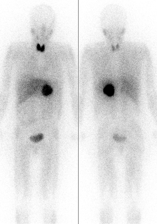
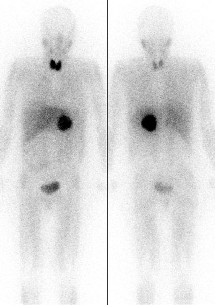
Pheochromocytoma (dark circular shadow near body center) localized by MIBG scintigraphy. Front and back views also show radioiodine collection in thyroid (neck) and bladder (pelvis). Source: Wikipedia
File history
Click on a date/time to view the file as it appeared at that time.
| Date/Time | Thumbnail | Dimensions | User | Comment | |
|---|---|---|---|---|---|
| current | 18:14, 13 March 2024 | | 526 × 747 (244 KB) | Urology4all (talk | contribs) |
You cannot overwrite this file.
File usage
The following page uses this file: